
The patient is a 50-year-old man who presents to the ED (emergency department) because of "palpitations". His Lead II Rhythm Strip is shown in Figure 1. The patient has a history of hypertension and is on a diuretic — but he is otherwise healthy. BP = 160/90 mmHg at the time the rhythm strip in Figure 1 is recorded. He has "heart awareness" (which is understandable given his rapid rate) — but otherwise no chest pain and no significant dyspnea.
- Is the patient in an SVT? IF so — Which one?
- How to proceed clinically?
- Should you give Adenosine? If you do and it works — Does this prove that the rhythm in Figure 1 is an SVT?
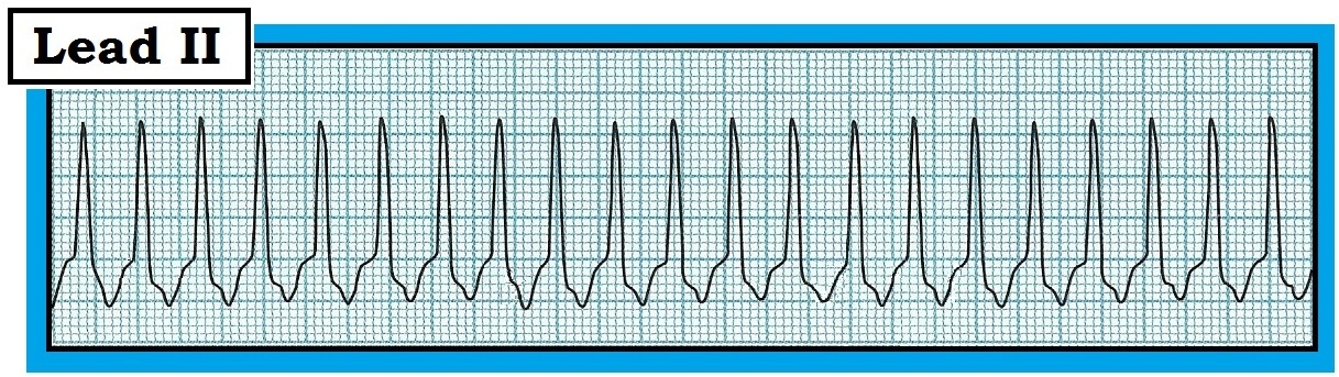 |
| Figure 1: Lead II rhythm strip obtained on a 50-year-old man with palpitations. BP = 160/90. Is this an SVT? How should you proceed clinically? - NOTE - Enlarge by clicking on Figures - Right-Click to open in a separate window. |
ANSWER: As is the case for clinical evaluation and management of any cardiac arrhythmia — the 1st thing to do is assess the patient!
- Ensure that the patient is hemodynamically stable! IF the patient is not stable as a direct result of the rapid rate — then electrical therapy (synchronized cardioversion or defibrillation) becomes immediately indicated regardless of whether the rhythm is VT, SVT, or WCT of uncertain etiology.
The patient in this case clearly appears to be stable. He is alert — having ‘heart awareness’ but no significant chest pain or dyspnea – and – his BP = 160/90 mmHg. Given that this patient is stable — we next assess the rhythm in Figure-1 by looking for the 5 KEY Parameters (“Watch your Ps and Qs – and the 3Rs" ):
- P Waves? — No P waves are seen (and no sign of atrial activity).
- QRS width? — Using the definition that a QRS duration of more than half a large box (>0.10 second) is “wide” — the QRS does not appear to be wide in the lead II rhythm strip shown in Figure-1.
- Regularity? — The overall rhythm in Figure-1 appears to be regular.
- Rate? — We estimate the rate in Figure-1 at just over 200/minute (By the “every-other-beat” method — the R-R interval of 2 beats is just under 3 large boxes; therefore ‘half’ the rate is just over 100/minute X 2 = ~210/minute for the estimated rate).
- Related? — There is no relation between P waves and the QRS (because there are no P waves to be seen … ).
WHAT Should Be Done at this Point?
- HINT: — Do we know for sure IF the QRS in Figure 1 is wide?
Points to emphasize from this case thus far include the following:
- The patient is stable — so by definition, we have at least a moment of time to contemplate management (remaining ever ready to cardiovert the patient IF at any time he becomes unstable).
- We do not yet know what the rhythm in Figure-1 is. All we know — is that this patient is in a stable Tachycardia (with a regular rhythm but no sign of atrial activity and a QRS complex that does not appear to be wide in the single lead that we see).
- We could reasonably treat the patient with Adenosine at this point.
- We should obtain a 12-lead ECG during the tachycardia as soon as it is possible to do so.
For conciseness — We limit illustration to a simultaneously recorded 2-lead rhythm strip of lead II and lead V1 (Figure-2).
- Do you still think the QRS complex in Figure-1 is narrow? How will the answer to this question affect your clinical approach?
 |
| Figure-2: Simultaneously-recorded rhythm strip of leads II and V1. Do you still think this patient is in a narrow-complex tachycardia? |
ANSWER to Figure 2: “12 Leads are Better than One”
It should be obvious that the QRS complex in Figure-2 is wide. The reason we initially thought the QRS in the lead II rhythm strip shown in Figure-1 was narrow — is that we misinterpreted the terminal part of the QRS as the beginning of the ST segment (See vertical red time lines in Figure-2). The rest of the 12-lead ECG obtained during tachycardia on this patient confirmed obvious QRS widening in virtually all leads except lead II. We emphasize the following points:
- The reason we were able to take the time to obtain additional leads on this patient — is that he was hemodynamically stable and tolerating the tachycardia in Figure-1. IF the patient was not stable (or at any time became unstable) — synchronized cardioversion would have been immediately indicated. Some patients may remain stable despite being in Tachycardia for an extended period of time (Cases of documented VT that persist for hours or longer are not uncommon). Knowing this — and knowing what to do IF at any time in the process the patient decompensates — affords the emergency care provider the luxury of at least a moment of time to contemplate further management.
- Optimal Management of Tachycardia depends on accurate Rhythm Diagnosis. This is not to say that management can't proceed before a definite diagnosis of the rhythm is made — but only that optimal management is best undertaken when the likely rhythm diagnosis is known.
- Once you have ensured that the patient with Tachycardia is stable — Use of the 5 KEY Parameters facilitates rhythm diagnosis (and allows you to narrow your differential even if unable to be absolutely certain of the rhythm). Therefore — “Watch your Ps and Qs — and the 3Rs” (Look for P waves or atrial activity — assess QRS width — determine Rate and Regularity of the rhythm — and if P waves are present, assess the Relationship [if any] between P waves and the QRS). Note: It does not matter in what sequence you look for these 5 parameters — as long as you always look for all 5 of them in every rhythm you encounter (We often alter the sequence we use depending on which parameters are easiest to identify for the rhythm at hand).
- Determining QRS Width is a KEY component for assessing Tachycardia. Remember — “12 Leads are Better than One”. Part of the QRS may lie on the baseline in the single monitoring lead being used (as was the case in Figure-1). IF the patient in Tachycardia is stable — Obtaining a 12-lead ECG during Tachycardia will often be revealing (and clarify IF the QRS is wide — as well as whether or not atrial activity is present).
BOTTOM LINE: Figure-1 is Really a Regular Wide Tachycardia!
The 2-lead rhythm strip shown in Figure-2 confirms that the 50-year-old man in this case is actually in a stable WCT rhythm (Wide-Complex Tachycardia).
- VT (Ventricular Tachycardia) must be assumed for the rhythm in Figure-1 until proven otherwise. This case brings home the point that “SVT” can not be diagnosed until one is certain about QRS width – and – one can not be certain the QRS is narrow until all 12 leads are seen to be narrow on a 12-lead ECG obtained during the tachycardia.
- Adenosine can still be given for the rhythm in Figure-1, even after we realize that the QRS is wide. This is because between 5-10% of VT rhythms are adenosine-responsive (See References below). That said — Once we realize that the QRS is WIDE (and that VT is far more likely than SVT) — our treatment priorities change to management of the regular, stable Wide-QRS Tachycardia).
-----------------------------------------------------------------------
-  -
-
Please Check out the following relevant PDF Sections excerpted from ACLS-2013-ePub:- Section 06.0 — on Using Adenosine -
- Section 07.0 — on Known VT -
- Sections 08.0, 09.0 — on The Regular Wide Tachycardia -
- Section 13.0 — on SVT of Uncertain Etiology -
-----------------------------------------------------------------------
No comments:
Post a Comment