Saturday, May 29, 2021

ECG Blog #229 — VT or Something Else?


The ECG shown in Figure-1 was obtained from an elderly man, who was admitted to the hospital with presumed Covid pneumonia. In addition to his pulmonary symptoms — he complained of palpitations and chest pain. No prior tracing was available. The patient was hemodynamically stable at the time ECG #1 was recorded.

  • WHAT is the rhythm? 
  • Is this VT — or something else?


Figure-1: ECG obtained from an elderly man with chest pain, palpitations and Covid pneumonia. What is the rhythm?


MThoughts on ECG #1:

I found this case challenging. It was sent to me without the benefit of any prior ECG for comparison. Below are "My Thoughts— that I review in the sequence that they came to me:

  • The rhythm in Figure-1 is a regular WCT (Wide-Complex Tachycardia) at a rate of ~150/minute — but without clear sign of sinus P waves. This immediately raises the possibility of VT (Ventricular Tachycardia). That said — since the patient is hemodynamically stable, we have the "luxury" of an additional moment to look further at the tracing.
  • Despite awareness that at least 80% of all regular WCT rhythms without sinus P waves will turn out to be VT — QRS morphology in Figure-1 really does look supraventricular! This is because: i) There is a very typical RBBB morphology in lead V1, with a triphasic rSR' in which the S wave descends below the baseline and there is a taller "right rabbit ear" (ie, a slender and taller R' complex); andii) A prominent R wave with fairly rapid initial upstroke and terminal S wave is seen in lateral leads I and V6. (NOTE: For review of these concepts — See ECG Blog #196 and ECG Blog #211).
  • I suspected that I was seeing atrial activity in several leads — possibly in the form of 2:1 AV conduction. 


COMMENT: I'll emphasize that it should take the experienced provider less than 20 seconds to arrive at the above impressions. 

  • I'll also emphasize that at this point — I was not at all certain about the etiology of the rhythm in Figure-1. That said — My "internal" estimation for the likely diagnosis of this WCT rhythm had moved from over 80% likelihood of VT — to a much greater probability that the rhythm was some type of SVT (SupraVentricular Tachycardia) because: i) QRS morphology looked so typical for RBBB conduction; and, ii) I really thought I was seeing regular underlying atrial activity in a number of leads.
  • CAVEAT #1: There are a number of forms of Fascicular VT, in which the QRS is not overly wide — and, QRS morphology may resemble RBBB conduction. Most of the time with Fascicular VT — there is a bifascicular block pattern (which we do not see here) — and there are also some slight differences from "typical" RBBB conduction (which I do not see here) — but because of this possibility of Fascicular VT, I was not yet 100% certain of a supraventricular etiology (For more on Idiopathic VT, including Fascicular VT — See ECG Blog #197).


MThoughts Continued on ECG #1:

Given that this patient was hemodynamically stable — I wanted to see IF I could "walk out" regular atrial activity (Figure-2):

  • PEARL #1: IF I was correct that the rhythm in Figure-1 was supraventricular — then description of our assessment to this point would be that of a regular SVT rhythm without clear sign of sinus P waves, at a rate of ~150/minute. Although possible that the rhythm could represent some form of "reentry" SVT (ie, AVNRT or AVRT) — this description should immediately prompt the diagnosis of AFlutter (Atrial Flutter) until proven otherwise. Knowing the likelihood of AFlutter should facilitate our search in Figure-2 for underlying atrial activity.
  • PEARL #2: Although my search for underlying (and partially hidden) atrial activity always encompasses assessment of all 12 leads — the leads I have found to be most helpful in this search for hidden atrial activity are: i) The inferior leads (surprisingly — lead III is often more helpful than lead II in finding non-sinus atrial activity!)ii) Lead aVRandiii) Leads V1 and V2.
  • Short, vertical RED lines in Figure-2 reveal strong suggestion that there is in fact regular underlying atrial activity — with 2 P waves occurring within each R-R interval in a number of leads. That this atrial activity is being conducted — is evident from the finding that the distance (ie, PR interval) from QRS complexes in selected leads to preceding atrial activity is constant — which means that there is 2:1 AV conduction.
  • PEARL #3: Although we can verify precisely regular 2:1 atrial activity with use of calipers — I'll emphasize that with practice, experienced providers can learn to instantly recognize these P-QRS relationships at the bedside (without a need to have to reach for calipers in front of an acutely ill patient).
  • PEARL #4: Because of suboptimal resolution, as well as some slanting of the ECG paper in Figure-2 — I've drawn in BLACK the outline of several large boxes on the ECG grid paper. Since the P-P interval of the vertical RED lines in this figure is approximately 1 large box in duration — this confirms that the atrial rate is ~300/minute — and the only rhythm that produces regular atrial activity at ~300/minute is Atrial Flutter!
  • CAVEAT #2: I am aware of rare cases of coexistent AFlutter and VT. That said — the combination of regular atrial activity at ~300/minute with 2:1 AV conduction + QRS morphology highly typical for RBBB conduction — strongly suggests that the rhythm in Figure-2 is indeed AFlutter in a patient with either preexisting RBBB or AFlutter with rate-related aberrant conduction. Clinically — since this patient was hemodynamically stable — I would have most probably begun rate-slowing medication on the assumption that the rhythm was AFlutter with RBBB conduction.


Figure-2: I've added vertical RED lines in selected leads to highlight regular underlying atrial activity. For clarity — I've oulined in BLACK the dimensions of a few large boxes, showing that the P-P interval is approximately 1 large box in duration (See text).


=======================================

NOTE: Today's 10:00 minute ECG Audio PEARL is all about Atrial Flutter.

=======================================


Today’s ECG Media PEARL #45 (10:00 minutes Audio) — Why is Atrial FIutter so commonly overlooked? Reviews PEARLS regarding the ECG diagnosis of AFlutter — and — What's "New"? in the field, regarding distinction between AFlutter vs Atrial Tachycardia (5/29/2021).

  • For those wanting a more advanced review on some newer concepts regarding AFlutter — Check Out this article by García-Cosío F et al (Clinical Approach to ATach and AFlutter, Rev Esp Cardiol 65(4):363-375, 2012).
  • For more on distinction between AFlutter vs ATach — Check out Figure-4 in the Addendum below.


The Case Continues:

The rapid rhythm in Figure-2 was treated with a beta-blocker. The patient's symptoms decreased — and a follow-up ECG was obtained (Figure-3).

  • WHAT does ECG #2 tell you about what the rhythm in ECG #1 was?
  • Is there anything else in ECG #2 to be concerned about?


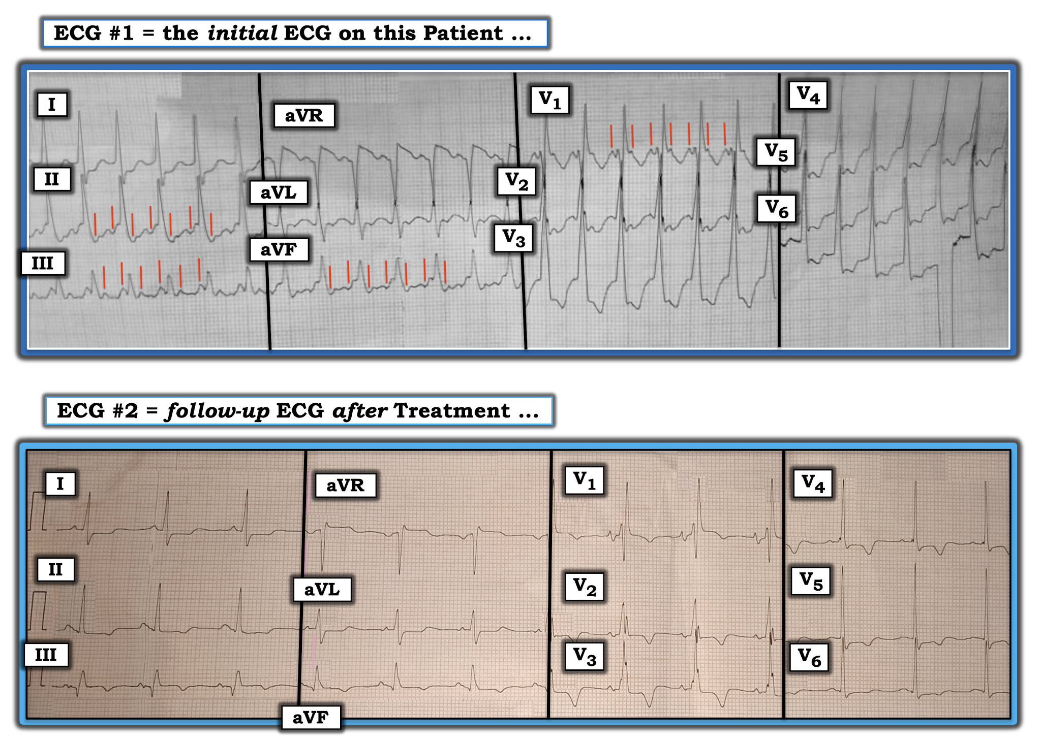
Figure-3: Comparison of the initial ECG with the 12-lead tracing obtained after treatment of the fast rhythm (See text).


MThoughts on Figure-3:

The "good news" — is that beta-blocker treatment successfully converted the tachycardia that was seen in ECG #1 to normal sinus rhythm (Note return of normal, upright sinus P waves, with fixed and normal PR interval in lead II of ECG #2).

  • PEARL #5: The post-conversion tracing may prove invaluable for confirming the etiology of an unknown tachycardia. Proof that the rhythm in ECG #1 was supraventricular — is forthcoming from the presence of identical QRS morphology in leads IV1 and V6 of ECG #2. Note the very typical RBBB morphology after conversion to sinus rhythm, with triphasic rSR' complex in lead V1 of ECG #2 (ie, with triphasic rSR' in which the S wave descends below the baseline and in which there is a taller "right rabbit ear" with a slender and taller R' complex). Lateral leads I and V6 manifest terminal S waves (as they should) in ECG #2 after conversion to sinus rhythm.
  • Proof that the vertical RED lines in ECG #1 truly represented 2:1 atrial activity — is forthcoming from the fact that the deflections highlighted by RED ines in ECG #1 are no longer present in the baseline after conversion to sinus rhythm.


PEARL #6: The post-conversion tracing may also provide insight into the occurrence of acute ECG changes that might not have been evident during the tachycardia.

  • A large Q wave is seen in lead III of ECG #2, in association with slight-but-real ST elevation and beginning T wave inversion in this lead.
  • A smaller but-definitely-present Q wave is also seen in lead aVF of ECG #2, in association with flattening (albeit no elevation) of the ST segment in this lead, with beginning T wave inversion.
  • Leads III and aVL all show similar ST segment straightening, with slight ST depression. 
  • Of note — the shape of the ST-T wave in lead aVL is the mirror-image opposite of the slightly elevated ST-T wave in lead III (See ECG Blog #184 for the potential significance of this finding).
  • PEARL #7: There is subtle-but-real ST elevation in lead V1. This is not normal — as the expected response for the ST-T wave in lead V1 with uncomplicated RBBB is if anything, slight ST-T wave depression (See ECG Blog #204 for the expected ST-T wave appearance with BBB)Proof that the ST-T wave response in the other chest leads of ECG #2 is not normal — is forthcoming from the finding that instead of the amount of T wave inversion decreasing (as it should) as one moves from leads V1-to-V2-to-V3 — there is increased deeping of T wave inversion (which is maximal in lead V3) — and which persists with 1-2 mm of ST depression through to lead V6.


Putting It All Together:

Today's case provides an excellent example of a regular WCT rhythm that we suspected was supraventricular from the initial ECG — and, which we were able to prove was supraventricular by comparison with the post-conversion tracing.

  • Focus on QRS morphology, with targeted search for hidden underlying atrial activity allowed us to quickly suspect a supraventricular etiology. Recognition of 2:1 atrial activity (in Figure-2) — with estimated atrial and ventricular rates of 300 and 150/minute, respectively — told us the rhythm was AFlutter within less than a minute.
  • Despite improvement in the patient's symptoms after conversion to sinus rhythm — review of the post-conversion 12-lead tracing strongly suggested ischemia at-the-least — and, most likely the presence of reperfusion ST-T wave changes secondary to a recent infarction (See ECG Blog #194  for review of ECG changes of coronary reperfusion).

ADDENDUM (June 3, 2021): I've been asked whether the post-conversion tracing in Figure-3 might raise the possibility of acute PE (Pulmonary Embolism) as the cause of this patient's acute pulmonary symptoms.
  • There is an S1Q3T3 pattern in ECG #2 ( = the post-conversion tracing) — but the S1, as well as the T3 are often "part" of the RBBB, which for all we know, may be a longterm finding in this patient (No prior tracing was available).
  • Other findings on ECG #2 could clearly be consistent with acute/recent PE (inferior and anterior ST-T wave changes that could reflect RV "strain" — and subtle ST elevation in right-sided leads III, aVR, V1).
  • The patient is no longer tachycardia in the post-conversion tracing (as might be expected if the cause of AFlutter was a large enough acute PE to produce both inferior and anterior ST-T wave changes of RV "strain") — BUT — use of the beta-blocker as treatment of the tachycardia (? the dose and duration of effect of that medication) may have attenuated a rapid heart rate.
  • Given the above — HOW to proceed would seem to depend on one's clinical suspicion for acute/recent PE (ie, I wouldn't expect all of this patient's symptoms to resolve after conversion to sinus rhythm if the cause of AFlutter was a large PE ... — but underlying Covid pneumonia clearly put this patient at increased risk for PE, and his post-conversion ECG could clearly be consistent with this diagnosis). Bottom Line: I agree that acute/recent PE should be part of the differential diagnosis of potential causes for this patient's episode of AFlutter. Wish we had more follow-up on this patient.


==================================

Acknowledgment: My appreciation to Drs. Chamanti and Anil Kumar Kolli (from Indiafor the case and this tracing.

==================================


ADDENDUM (May 29, 2021): I've excerpted from my ACLS-2013-ePub this brief section that reviews some subtleties to consider when contemplating a differential diagnosis between AFlutter vs ATach (Figure-4).


Figure-4: Distinction between AFlutter vs ATach (More on this in today's Audio Pearl above).


================================== 


Related ECG Blog Posts to Today’s Case: 

  • ECG Blog #185 — Reviews the Ps, Qs & 3R Approach to Systematic Rhythm Interpretation
  • ECG Blog #196 — Reviews "My Take" on assessing the regular WCT (Wide-Complex Tachycardia) — with tips for distinguishing between VT vs SVT with either preexisting BBB or aberrant conduction.
  • ECG Blog #211 — Reviews WHY some early beats and some SVT rhythms are conducted with Aberration (and why the most common form of aberrant conduction manifests RBBB morphology).
  • ECG Blog #197 — Reviews the common forms of Idiopathic VT (including Fascicular VT). 
  • ECG Blog #184 — Reviews the "Magical" Lead 3-Lead aVL Relationship with acute OMI (Occlusion-based Myocardial Infarction).
  • ECG Blog #204 Reviews my "user-friendly" approach for the ECG diagnosis of BBB (including assessment of expected ST-T wave changes with RBBB and LBBB)
  • ECG Blog #194 — Reviews how to tell IF the "culprit" artery has reperfused (using clinical and ECG criteria).


Wednesday, May 26, 2021

ECG Blog #228 (44) — What is the Main Problem?


The 12-lead ECG and long lead II rhythm strip shown in Figure-1 was obtained from a middle-aged woman with a several day history of fever and shortness of breath. She had palpitations — but NO chest pain. The patient tested positive for Covid-19 several days earlier.

  • How would YOU interpret this tracing?
  • What is the main problem?


Figure-1: ECG obtained from a middle-aged woman with fever, dyspnea, palpitations, and a positive test for Covid-19. NO chest pain.


=======================================

NOTE #1: Some readers may prefer at this point to listen to the 3:30 minute ECG Audio PEARL before reading My Thoughts regarding the ECG in Figure-1. Feel free at any time to review to My Thoughts on this tracing (that appear below ECG MP-44).

=======================================


Today’s ECG Media PEARL #44 (3:30 minutes Audio) — What is a "Silent" MI? — and How OFTEN do "Silent MIs" occur? (results from Framingham — 5/24/2021).


MY Initial Approach to the ECG in Figure-1:

Unfortunately, the tracing in Figure-1 is curved — therefore a bit distorted (was sent to me via the internet). That said — this ECG is of good enough quality to appreciate the important ECG findings.

  • The rhythm is venricular bigeminy — that is, every-other-beat ( = beats #1, 3, 5, 7, 9, 11, 13 and 15) is a PVC (Premature Ventricular Contraction). We know these beats are PVCs — because the QRS complex is wide; it looks very different than the sinus-conducted beats; and the QRS of these wide beats is not preceded by a P wave.


PEARL #1: To determine what else might be ongoing in this ECG — We need to focus on ST-T wave morphology of sinus-conducted beats in all 12-leads (ie, for beats #2, 4, 6, 8, 10, 12 and 14)

  • Beyond-the-Core: There are 2 possible exceptions to the statement I just made, namely that, "We need to focus on ST-T wave morphology of sinus-conducted beats". It is uncommon that either of these exceptions will apply — but for advanced interpreters, it is worthwhile being aware of them: i) Ventricular ectopy itself (not necessarily related to acute or longterm coronary disease) may alter the ST-T wave of the sinus-conducted beat that follows a PVC; andii) On occasion, it is the ST-T wave of a PVC that provides the best indication of acute coronary disease (ie, when the PVC shows obviously abnormal ST elevation or depression).


Because of the large size and frequency of the PVCs in Figure-1 — it might be EASY to overlook the abnormal ST-T wave findings of the sinus-conducted beats. 

  • To facilitate this assessment — I have enclosed the QRS complex and ST-T wave of each of the sinus-conducted beats in all 12 leads within a BLUE-WHITE rectangle (Figure-2).


Figure-2: I've enclosed the QRS complex and ST-T wave from all sinus-conducted beats within one of the BLUE-WHITE rectangles (See text).


MY Interpretation of Figure-2:

QRS amplitude for each of the 6 limb leads is small, especially in comparison to the much larger amplitude of the PVCs.

  • There is ST segment coving and slight-but-real ST elevation in each of the 3 inferior leads (II, III, aVF). ST elevation is most marked in lead III — and the "takeoff" of the ST segment in all 3 inferior leads is abnormally straightened (slanted BLUE lines drawn parallel to the ST segment "takeoff" in these 3 leads).
  • Confirmation of recent or acute inferior infarction is forthcoming from the finding of reciprocal ST depressionin high-lateral leads I and aVL.
  • Associated posterior MI is diagnosed by: i) Early transition, with a predominantly positive R wave already in lead V2; andii) Shelf-like ST depression in the anterior leads, that is maximal in lead V3.
  • There is also ST-T wave flattening with slight depression in the lateral chest leads (V4, V5 and V6).


Putting It All Together:

This middle-aged woman with fever, dyspnea with Covid-19 positivity and palpitations — but no chest pain— presented to the ED with a very abnormal ECG:

  • The rhythm was ventricular bigeminy.
  • Assessment of QRST morphology of sinus-conducted beats (Figure-2) — suggested recent (if not acute) infero-postero MI.
  • The reason for the frequent PVCs is most probably the recent acute MI.
  • PEARL #2: This patient had a number of acute symptoms — but no chest pain. This satisfies the definition of a "silent" MI (which is the topic of our brief Audio Pearl #44See above!)


The Case Continued:

A follow-up ECG was obtained on this patient (Figure-3). Now that the PVCs have resolved in ECG #2 — Isn't it easier to identify the acute findings on this tracing!

  • QUESTION: What is the probable "culprit" artery?


Figure-3: Follow-up ECG obtained in today's case (See text).


ANSWER:

The rhythm in ECG #2 (shown in Figure-3) is sinus at ~75/minute. All intervals are normal. There is slight axis deviation — but not enough to qualify as LAHB (ie, the QRS is still predominantly positive in lead II). There is no chamber enlargement.

  • A large Q wave is now seen in lead III (A small-but-present initial r wave was seen in lead III in Figure-2).
  • R wave progression in Figure-3 is abnormal — with early transition and a predominant R wave already in lead V2.
  • Although QRS amplitude in the limb leads remains small — there appears to be more ST elevation in inferior leads III and aVF than there had been in Figure-2.
  • The ST depression in lead aVL is also increased compared to Figure-2 — and now takes on a "magical" mirror-image opposite picture, compared to the shape of the ST elevation in lead III (See ECG Blog #184 for more on this "magical" mirror-image opposite picture between leads III and aVL in the setting of acute infarction).
  • In the chest leads — We have already noted the early transition with a surprisingly tall R wave already in lead V2. This is associated with marked ST depression in lead V2, with terminal positivity of the T wave. The ECG appearance of the QRST complex in lead V2 constitutes a positive "Mirror Test" — that is virtually diagnostic of acute posterior MI (See ECG Blog #80 and ECG Blog #56). But what is most remarkable about the ST-T waves in the chest leads of Figure-3 — is that despite marked ST depression in lead V2, there is virtually no ST depression in lead V1. This suggests the strong possibility of associated acute RV (Right Ventricular) infarction (See ECG Blog #190-LINK).
  • PEARL #3: The probable "culprit" artery in today's case is the RCA (Right Coronary Artery) because: iAcute infero-postero MI in the absence of anterior ST elevation narrows down the possibilities for the "culprit" artery to the RCA or the LCx (Left Circumflex) coronary artery — and 80-90% of individuals have a right-dominant circulation (ie, statistical odds favor the RCA); ii) The finding of more ST elevation in lead III > II — together with marked, mirror-image opposite reciprocal ST depression in lead aVL favors the RCA; andiiiThe flat ST segment in lead V1, in association with marked ST depression in lead V2 suggests acute RV involvement — and the LCx does not vascularize the right ventricle.


Case Follow-Up:

The acute MI was recognized by the treating clinicians. Since acute cath facilities were not available — the patient was treated with thrombolytic therapy.


==================================

Acknowledgment: My appreciation to Drs. Chamanti and Anil Kumar Kolli (from Indiafor the case and this tracing.

==================================


Related ECG Blog Posts to Today’s Case: 

  • ECG Blog #184 — Reviews the "Magical" Lead 3-Lead aVL Relationship with acute OMI (Occlusion-based Myocardial Infarction). 
  • ECG Blog #80 — Reviews the positive "Mirror Test" for ECG diagnosis of acute posterior MI — as well as the basics for predicting the "culprit" artery with acute MI. 
  • ECG Blog #56 — Reviews the positive "Mirror Test" for ECG diagnosis of acute posterior MI
  • ECG Blog #190 — Reviews the ECG diagnosis of acute RV (Right Ventricular) MI
  • ECG Blog #193 — Reviews the concept of "OMI" ( = Occlusion-based Myocardial Infarction) — with some basics on the "Mirror Test" — and for predicting the "culprit" artery plus links to PDF summaries and numerous other acute MI cases from my ECG Blog.


Sunday, May 23, 2021

ECG Blog #227 — Sinus Tach and 1st-Degree?


The ECG in today's case was obtained from a middle-aged man with a 2-day history of exertional chest pain and shortness of breath (Figure-1). Questions raised by this case include the following:

  • Is the rhythm sinus tachycardia with 1st-degree AV block?
  • Is there evidence of an inferior MI? 
  • Are Sgarbossa criteria applicable to this tracing?


Figure-1: ECG obtained from a middle-aged man with exertional chest pain and shortness of breath. The rhythm was thought to be sinus tachycardia with 1st-degree AV block (RED arrow in lead II highlighting the upright sinus P wave).


=======================================

NOTE: Unfortunately I do not have follow-up to this case — so I do not know a definitive answer regarding the cardiac rhythm. That said — I believe there are important lessons-to-be-learned with respect to the diagnostic considerations that should immediately come to mind (!) — and THAT is the purpose of today's case.

  • HINT: Today's 1:45 minute ECG Audio PEARL #43 (below) reviews an important arrhythmia concept known as the Bix Rule.

=======================================


Today’s ECG Media PEARL #43 (1:45 minutes Audio) — Reviews the Bix Rule (which explains why it is uncommon to see sinus tachycarda with 1st-degree AV block).


My THOUGHTS on ECG #1:

My initial impression on assessing the ECG rhythm in Figure #1 was sinus tachycardia with 1st-degree AV block. And then I took a 2nd look ...

  • PEARL #1: As the rate of a sinus rhythm increases — the PR interval tends to shorten, probably as a result of reduced basal vagal tone. Because of this, it is uncommon to see sinus tachycardia with a prolonged PR interval. While there clearly are some patients who have baseline PR interval prolongation, who continue to manifest a certain amount of PR interval prolongation with sinus tachycardia — a majority of tachycardias that seem to show a "sinus P wave" in the middle of the R-R interval turn out to have 2:1 AV conduction (from either Atrial Tach or AFlutter). This principle is known as the Bix Rule, named after the Viennese cardiologist who first cautioned against "accepting" the diagnosis of "sinus tach with 1st-degree" before meticulous search for an "extra" P wave hidden within the QRS complex.


Acknowledgment: It is unfortunately impossible to prove that there is 2:1 AV conduction in Figure-1 — without either a prior baseline tracing showing identical QRS morphology with definite sinus rhythm at a slower rate — and/or — without a follow-up tracing showing the same QRS morphology and definite sinus rhythm at a slower rate.

  • That said, I thought: i) The PR interval for the upright deflection in Figure-1 looked "long" given the tachycardia rate; andii) Each of the inferior leads showed a potential "hump" or deflection that might represent a near-simultaneous "extra" P wave in association with the end of the QRS complex — that seemed to "walk out" with calipers (short RED lines in the inferior leads in Figure-2).
  • In lead V1 — we clearly see a negative P wave deflection before each QRS complex — which if "walked out" with calipers, could easily "hide" a second P wave within the QRS complex (short RED lines in lead V1 of Figure-2).
  • PEARL #2: One of the most commonly overlooked arrhythmia findings is subtle 2:1 AV conduction. Over the years — I have seen too-numerous-to-count experienced providers (including many cardiologists) overlook 2:1 AV conduction with either AFlutter or ATach. The BEST way not to overlook subtle 2:1 AV conduction — is to THINK of this possibility with fast supraventricular rhythms, in which you either do not see a clear upright P wave in lead II — or — in which the PR interval looks "longer-than-expected" for a normal sinus P wave ( = the Bix Rule!)NOTE: I fully acknowledge that 2:1 conduction will not be present each time you look for it (!) — but you will not pick up most subtle 2:1 conduction rhythms unless you regularly look for them!


What Happened in Today's Case:

I believe the possibility of 2:1 AV conduction was not considered in today's case — so unfortunately, I do not know "the Answer".

  • IF indeed, the rhythm in Figure-2 was sinus tachycardia — then there would be no need to "treat the rhythm". Instead — the treatment of choice for sinus tachycardia is to treat the underlying cause (in this case, whatever was causing this patient's exertional chest pain and shortness of breath) — in which case, the rate of sinus tachycardia should slow as the patient's clinical condition improves, and clear definition of the rhythm will then most probably be forthcoming!
  • Alternatively — another lead system (such as use of a Lewis Lead) might be tried in an attempt to better visualize atrial activity (See Figure-3 below in the Addendum).


Figure-2: I've added short RED lines to Figure-1 at places where I suspected there might be underlying P waves with 2:1 AV conduction (See text).


Other ECG Findings in Figure-2:

  • Although the QRS complex in Figure-2 looks to be a little bit wide (ie, slightly more than half a large box in duration) — it does not look wide enough by my calculation to qualify as a complete LBBB (ie, not as wide as ≥0.12 second in duration).
  • In addition to not being overly wide — QRS morphology in Figure-2 "looks" supraventricular. Note how QRS amplitude is dramatically increased! For clarity (because of the marked overlap in complexes in Figure-2) — I have outlined in PURPLE the limits of the QRS in lead V3 — and in PINK, the limits of the QRS in lead V5. Both of these QRS complexes are cut off at their peak, but already measure more than 25 mm!
  • PEARL #3: The precise cause of slight QRS widening in this case is difficult to determine. I suspect there is a combination of marked LVH (a thicker left ventricle takes up to 0.01-to-0.02 seconds more to traverse) and possible incomplete LBBB.To my knowledge — there is no literature validation on use of Sgarbossa criteria for incomplete LBBB (unlike complete LBBB — incomplete LBBB is too uncommon to objectively study). That said — common sense would suggest that the rule of proportionality conveyed by the Smith modification of Sgarbossa criteria should still apply. I see no abnormal-looking ST elevation in Figure-2. And although the amount of J-point ST depression in lateral chest leads V4-thru-V6 is marked (especially in lead V4) — given huge R wave amplitude in these leads, my "sense" is that J-point ST depression is probably not "disproportionate" given how huge R wave amplitude is.
  • As to the question of possible inferior MI of uncertain age — an isolated inferior Q wave in lead III is not necessarily abnormal, especially in association of possible incomplete LBBB.


Putting It All Together:

The ECG in today's case shows a supraventricular rhythm at a rate just over 100/minute. The QRS complex appears to be slightly wide, albeit with a supraventricular-like morphology most consistent with marked LVHand possible incomplete LBBB.

  • The cardiac rhythm is uncertain. The rhythm could be sinus tachycardia with 1st-degree AV block — or — this could be atrial tachycardia with 2:1 AV conduction.
  • The "good news" — is that there do not appear to be acute ST-T wave changes on this ECG. One would "have to be there" in order to know how best to proceed. IF the patient was (and remained) hemodynamically stable — then treating associated disorders (ie, the cause of this patients exertional chest pain and shortness of breath) should result in a slowing of the rate if the rhythm was sinus tachycardia.
  • Finding a prior tracing — considering additional lead systems (such as use of a Lewis Lead) — and/or — repeat ECGs as the patient is treated would almost certainly clarify what the true cardiac rhythm is.


==================================

Acknowledgment: My appreciation to Abdullah Al Mamum (from Dhaka, Bangladesh) for allowing me to use this case and this tracing.

==================================


ADDENDUM (May 23, 2021): I've excerpted from my ACLS-2013-ePub directions on how to obtain a Lewis Lead, to help visualize atrial activity (as discussed in ECG Blog #223).


Figure-3: How to record a Lewis Lead.
 

================================== 

Related ECG Blog Posts to Today’s Case: 

  • ECB Blog #221 — Reviews my "user-friendly" approach for diagnosing the BBBs (Bundle Branch Blocksin less than 5 seconds — including Audio Pearl-38 on HOW to diagnose acute MI when there is BBB (with review of modified Smith-Sgarbossa criteria). 
  • ECG Blog #223 — Assessing P Waves and Atrial Activity (including use of a Lewis Lead).