Wednesday, October 27, 2021

ECG Blog #258 (70) — How to "Date" an MI?


The ECG shown in Figure-1 was obtained from an older patient with known coronary disease, who presented with a 10-hour history of worsening chest pain.

  • Given the above history — How would YOU interpret this tracing?
  • Should the cath lab be activated?
  • Extra Credit: Can you envision a clinical "story" to explain all ECG findings?
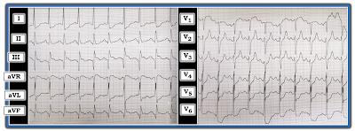

Figure-1: ECG obtained from an older patient with known coronary disease and chest pain of ~10 hours duration (See text).


=======================================

NOTE: Some readers may prefer at this point to listen to the 10:20-minute ECG Audio PEARL before reading My Thoughts regarding the ECG in Figure-1. Feel free at any time to refer to My Thoughts on this tracing (that appear below ECG MP-70).

=======================================


Today’s ECG Media PEARL #70 (10:20 minutes Audio) — Reviews HOW to "Date" an Infarction based on the initial ECG.


MY Sequential Thoughts on the ECG in Figure-1:

As always — I favor the use of a Systematic Approach (which I review in ECG Blog #205). Use of a systematic approach is especially important in today's case — because there are multiple ST-T wave abnormalities that otherwise might be easy to overlook.

  • Rate & Rhythm: The heart rate is fast. The P wave is upright in lead II, with a fixed and normal PR interval — so the rhythm in Figure-1 is sinus tachycardia at ~120-125/minute
  • Intervals (PR/QRS/QTc): The PR interval is normal — the QRS complex is narrow — and, the QTc may be slightly prolonged (albeit difficult to assess the QTc interval when the heart rate is faster than 90-100/minute).
  • Axis: The frontal plane QRS axis is normal (I'd estimate at about +20 degrees — given slightly more net positivity in lead I compared to lead II, with a near-isoelectric QRS in lead III).
  • Chamber Enlargement: The standardization marker is missing — so the best we can do is to assume normal standardization. Regardless of the standardization — I suspect there is LVH (Left Ventricular Hypertrophy) — given the tall R wave in lead I (which is cut off — but looks to be at least 15 mm) — and especially given the very tall R wave in lead V6 (of at least 20 mm — though as shown in Figure-2, precise measurement is difficult given overlap with the QRS in leads V4 and V5) — See ECG Blog #245 for details on LVH criteria.


Regarding Q-R-S-T Changes:

  • Q Waves — There are very large Q waves in 2 of the 3 inferior leads (ie, in leads III and aVF) — with a smaller-but-definitely-present Q wave in the 3rd inferior lead ( = lead II). T
  • R Wave Progression — Transition (where the R wave becomes taller than the S wave is deep) is appropriate, occurring between leads V3-to-V4. There may be a slight chest lead placement error for lead V2 (since the R wave and S wave in this lead are both larger than the R and S waves in both neighboring leads = V1 and V3, and normally that should not be). That said — even if present, this slight lead placement error should not alter the overall interpretation of this tracing.
  • ST-T Wave Changes — There is significant ST elevation of coved shape in lead III. There is abnormal ST elevation of a lesser degree (with a similar shape) in lead aVF. However, there is no ST elevation in the 3rd inferior lead (which is lead II). There is 1-2 mm of ST elevation in lead aVR.
  • T waves are surprisingly tall and peaked in leads V2 and V3, and to a lesser extent in lead V4. Unlike what occurs with hyperkalemia — there is no T wave peaking in any of the other 9 leads on this tracing.
  • There is marked ST depression in 5 of the remaining leads (ie, in leads I, aVL; V4, V5,V6).


Figure-2: I've labeled the ECG in today's case. Although overlap of the QRS in leads V4, V5 and V6 makes precise measurement of QRS amplitude difficult — the RED outline of the QRS in lead V6 suggests an R wave amplitude of at least 20 mm, which easily satisfies criteria for LVH. The inserts show application of the "Mirror Test" to illustrate the "magical" mirror-image opposite relationship with recent or acute infarction between leads III and aVL — and application of the "Mirror Test" in leads V2 and V3 suggests there are reperfusion T waves corresponding to recent posterior infarction (See text).


Putting IAll Together: There is a lot going on in this case. Although I lack details of follow-up for this patient — I nevertheless thought it worthwhile to work through what I feel is the most logical clinical scenario — as I believe the brief History we are given and this single ECG do tell a StoryBOTTOM LINE — I thought the ECG in Figure-1 strongly suggests there has been a recent (if not still acutely ongoing) infero-postero STEMI ( = ST Elevation Myocardial Infarction). I'll add the following thoughts:

  • The History we are given (namely that the patient is an older adult with known coronary disease — who presented with a 10-hour history of worsening chest pain) — is essential for optimal assessment and clinical decision-making.
  • Given this history and this patient’s ECG — there should be no doubt that inferior MI has occurred at some point in time. Large Q waves are seen in each of the 3 inferior leads (especially in leads III and aVF). That said — the clinical challenge is to determine, "What happened when?" — and — whether the cath lab should be immediately activated?
  • Of clear concern is the surprisingly fast rate of this patient's sinus tachycardia (ie, between 120-125/minute). Uncomplicated infarction would not be expected to cause a tachycardia this fast — which raises the concern that this patient may be decompensating (ie, pehaps in severe heart failure? — and/or with some other issue requiring immediate attention).


So HOW can we “date” this patient’s inferior MI? "Dating" an infarction is challenging! I address this question in the above Audio Pearl #70. There is conflicting evidence on this tracing with regard to the "age" of infarction.

  • The extremely large Q waves in leads III and aVF favor prior infarction. That said — it is possible that this older patient with known coronary disease had a previous inferior infarction — and is now having acute reinfarction of the inferior wall. PEARL: On occasion, significant Q waves have been known to form in as little as 1-to-2 hours. THEREFORE: We can not rule out the possibility of a very recent, or even acute ongoing infarction simply on the basis of the large size of these inferior Q waves.
  • There is coved ST elevation in lead III — and to a lesser extent in lead aVF. In addition — there is marked ST depression in no less than 5 leads (ie, leads I, aVL; V4, V5 and V6). This ST depression is clearly more marked and of a different "shape" than would be expected simply from LVH with "strain". I therefore interpreted this as reciprocal and/or ischemic ST depression from an acute (potentially ongoing) coronary event until proven otherwise.
  • In further support of a recent (if not acute) cardiac event — is the "magical" mirror-image opposite relationship of ST-T waves in leads III and aVL. As discussed in ECG Blog #184 and illustrated by the "mirror test" inserts I added for leads III and aVL in Figure-2 — the mirror-image of the ST-T wave depression in lead aVL looks identical to the ST elevation seen in lead III. Similarly — the mirror-image of the ST-T wave elevation in lead III looks identical to the shape of the ST depression seen in lead aVL.
  • That said — against the inferior infarction being acute is the fact that the amount of ST elevation in lead aVF is no more than minimal — and the complete lack of any ST elevation in lead II. Given the amount of ST depression in multiple leads on this tracing — I would have expected definite ST elevation of comparable amount in the inferior leads IF the infarction was acute.
  • A striking finding is the T wave peaking seen in leads V2 and V3. We see a continuation of this unexpectedly marked T wave peaking (following depression of the ST segment) in lead V4. This is not the T wave peaking of hyperkalemia — since it is only present in these 3 leads (and we’d expect more generalized T wave peaking if there was hyperkalemia). Instead — I feel the most logical explanation is illustrated by the "mirror test" inserts added in Figure-2 for leads V2 and V3 — which suggest that the mirror-image of the peaked T waves in leads V2, V3, V4 most likely represents deeply inverted reperfuson T waves from recent posterior infarction (this concept further reviewed in ECG Blog #193).
  • Finally — we are left with ST elevation in lead aVR — which, in association with the marked ST depression in 5 of the remaining leads (ie, leads I, aVL; V4,5,6) — raises concern for diffuse subendocardial ischemia resulting from significant coronary disease in other anatomic areas.


My SYNTHESIS (Explaining All ECG Findings in Today's Case):

The ECG findings in today's case can not be explained by a single acute event. Instead — I suspect the following:

  • This patient had a recent, large infero-postero STEMI. The limited amount of ST elevation in inferior leads — in association with surprisingly tall, peaked anterior T waves suggest there has been some spontaneous reperfusion (and that infarction occurred at least a number of hours ago — which is supported by the history stating that chest pain has been present for 10 hours).
  • This patient most probably has multi-vessel disease — with the marked ST depression in multiple leads potentially representing ischemia in other anatomic areas (and potentially attenuating ST elevation that otherwise may have been seen).
  • There is LVH — but the shape and amount of ST depression in lateral leads represents more than just LV "strain".
  • The marked sinus tachycardia (of ~120-125/minute) — in association with the above ECG findings and persistence of chest pain (worsening over 10 hours) — are clear indication for activating the cath lab! Even though there is ECG evidence for reperfusion — what has recently reopened might once again closeand — the marked sinus tachycardia + marked ST depression in multiple leads + persistent and worsening chest pain all argue for promptly defining the anatomy to determine IF there is need for acute PCI (Percutaneous Coronary Intervention).


============================== 

ADDENDUM (10/28/2021) — There are elements of this case that closely resemble Aslanger's Pattern (This pattern is very nicely described by Dr. Smith in his January 4, 2021 post). The premise of Aslanger's — is that IF there is inferior MI + diffuse subendocardial ischemia — then the vector of ST elevation will shift rightward. This results in:

  • ST elevation in lead III (as a result of the acute inferior MI) — but not in the other inferior leads (II, aVF) because of the rightward shift in the ST elevation vector.
  • ST depression in one or more of the lateral chest leads (V4, V5, V6) with a positive or terminally positive T wave — but without ST depression in lead V2. (Marked ST depression from multi-vessel coronary disease serves to attentuate what would have been ST elevation in leads II and aVF).
  • ST elevation in lead V1 that is more than any ST elevation in lead V2.
  • There may be more reciprocal ST depression in lead I than in lead aVL (because of the rightward ST vector shift).
  • The only leads showing significant ST elevation may be leads III, aVR and V1 (reflecting the inferior MI + subendocardial ischemia from diffuse coronary disease). 


The above said — additional features to consider in today's case include: i) There is also marked LVHandii) The infero-postero MI is probably at least 10 hours old, and is now showing prominent reperfusion T waves in the posterior distribution (ie, tall, peaked anterior T waves).


==============================

Acknowledgment: My appreciation to Ahmad Separham (from Tabriz, Iran) for the case and this tracing.

==================================


Related ECG Blog Posts to Today’s Case:

  • ECG Blog #205 — Reviews my Systematic Approach to 12-lead ECG Interpretation.

  • ECG Blog #245 — Reviews "My Take" on a user-friendly approach to the ECG diagnosis of LVH (including CAVEATS that may complicate assessment of associated acute ischemia).
  •  
  • ECG Blog #142 — Presents another case for discussion on how to "date" an infarction.

  • ECG Blog #193 — illustrates use of the Mirror Test to facilitate recognition of acute Posterior MI. This blog post reviews the basics for predicting the "culprit artery". NOTE: Figure-5 in the Addendum of this blog post illustrates the essentials for identifying an isolated posterior MI.

  • ECG Blog #184 — illustrates the "magical" mirror-image opposite relationship with acute ischemia between lead III and lead aVL (featured in Audio Pearl #2 in this blog post).

  • The September 21, 2020 post in Dr. Smith's ECG Blog — My Comment (at the bottom of the page) emphasizes utility of the Mirror Test for diagnosis of acute Posterior MI
  • The February 16, 2019 post in Dr. Smith's ECG Blog — My Comment (at the bottom of the page) emphasizes utility of the Mirror Test for diagnosis of acute Posterior MI
  • ECG Blog #80 — reviews prediction of the "culprit" artery (and provides another case illustrating the Mirror Test for diagnosis of acute Posterior MI).


Friday, October 15, 2021

ECG Blog #257 (19a) — AV Block in a Young Adult?


The 2-lead rhythm strip shown in Figure-1 was obtained from a young adult woman who presented for "palpitatons". No known previous history of heart disease.

  • How would YOU interpret this tracing?
  • Given the above history — What are your clinical considerations?


Figure-1: 2-lead rhythm strip obtained from a young adult woman with "palpitations" (See text).


=======================================

NOTE #1: Some readers may prefer at this point to listen to the 6:45-minute ECG Audio PEARL before reading My Thoughts regarding the ECG in Figure-1. Feel free at any time to refer to My Thoughts on this tracing (that appear below ECG MP-19a).

=======================================


Today’s ECG Media PEARL #19a (6:45 minutes Audio) — Reviews a few quick things to look at that allow you to rule in or rule out complete AV Block within seconds.


================================

NOTE #2: Although I lack clinical details for this case (and I don't even have access to a 12-lead ECG on this patient) — the 2-lead rhythm strip that we are provided in Figure-1, and the limited history we are given is still enough for an instructive discussion.

================================


My Sequential Thoughts for Interpreting this Tracing:

As always — I began my systematic approach to the rhythm with assessment of the PsQs and 3Rs (as discussed in detail in ECG Blog #185).

  • P waves — Sinus P waves are present in today's tracing! It was not immediately obvious to me that the atrial rhythm is regular — because many of the P waves either occur within the QRS complex or within the ST-T wave. In Figure-2 — I highlight with RED arrows a number of P waves that I can definitely identify.


Figure-2: I highlight a number of P waves from Figure-1 that I can definitely identify (RED arrows).


PEARL #1: This is the type of tracing for which use of calipers is essential! I know of no other way to determine that the atrial rhythm in Figure-2 is regular.

  • It is virtually impossible to determine the rhythm in Figure-2 unless you establish that the atrial rhythm is regular throughout the tracing.
  • It will literally take you less than 10 seconds to walk out regularly-occurring deflections (corresponding to regular P waves) with the use of calipers (RED arrows in Figure-3). 


Figure-3: Using calipers allows us to establish that the atrial rhythm is regular throughout today's tracing (RED arrows).


Continuing with the PsQs and 3R Approach:

  • The QRS complex in Figure-3 looks wide! (ie, about 3 little boxes = 0.12 second in duration). Unfortunately — we do not have access to a 12-lead ECG on this patient. That said — the predominantly negative morphology of the QRS complex in leads II and III is consistent with LAHB (Left Anterior HemiBlock).


PEARL #2 (Beyond-the-Core): Although the presence of a hemiblock may slightly prolong the process of ventricular depolarization — it usually does not do so by more than 0.01-to-0.02 second. Therefore — the wide QRS complex (that we measured in Figure-3 to be 0.12 second in duration) is longer than we would expect for simple LAHB. This suggests that there may be additional conduction system disease (ie, possible also RBBB = Right Bundle Branch Block).

  • While LAHB is a common ECG fnding in an older adult popultion — it is not commonly seen in otherwise healthy young adults. Therefore, even without seeing a complete 12-lead ECG on the young adult woman in today's can — we can suspect that there may be underlying structural (and/or conduction system) disease.


================================

Technical POINT: IF you look closely at the ECG grid lines in Figure-3 — it should be apparent that the ECG paper is angled (ie, the ECG grid boxes are not completely vertical — this being easiest to see at the beginning and end of the tracing). In addition, there is baseline artifact (more marked in lead II than lead III) — and — the ECG baseline is rising (being several boxes higher for the last few beats in the tracing). 

  • NOTE: While today's tracing is still adequate for interpretation — the above technical factors combine to produce slight distortion of measurements. The reason this is important — is that often the BEST clue that complete AV block is not present, is when one or more ventricular beats occur earlier-than-expected — which is why precise measurements are needed (This concept discussed in detail in today's Audio Pearl that appears above).


================================

Completing systematic assessment with the 3Rs:

  • Accounting for the above technical imperfections — I thought the ventricular rhythm in Figure-3 appeared to be Regular at a Rate of ~60/minute (ie, the R-R interval is ~5 large boxes in duration).
  • As per our caliper measurement (and as per the RED arrows in Figure-3) — the atrial rhythm in today's tracing appears to be Regular at the rapid atrial Rate of ~130/minute.
  • The 3rd "R" is "Related" — as determined by how many (if any) P waves in Figure-3 are related to neighboring QRS complexes.


PEARL #3: I find it easiest to determine if P waves are related to neighboring QRS complexes — by focusing on each QRS complex in the tracing, and looking to see if any PR intervals are constant.

  • In Figure-3 — the PR interval is constantly changing in front of each of the 10 beats on this tracing. This finding — plus the finding that both the atrial and ventricular rates in Figure-3 remain regular throughout the tracing — suggests that none of the P waves are being conducted to the ventrcles.
  • As emphasized earlier — the BEST clue that complete AV block is not present, occurs when one or more ventricular beats occur earlier-than-expected. This does not happen in Figure-3 — because the ventricular rate remains regular throughout the tracing!


Drawing the LADDERGRAM:

A picture tells 1,000 words. The Laddergram that I've drawn in Figure-4 illustrates the mechanism I propose for today's rhythm (See ECG Blog #188 for review on how to read and/or draw Laddergrams). The laddergram in Figure-4 suggests the following:

  • The atrial rhythm is rapid and regular — but none of the P waves are able to penetrate the AV node.
  • propose that the escape rhythm is junctional (regularly-occurring RED circles within the AV Nodal Tier). That said, the QRS complex is wide — and without a complete 12-lead ECG (and without a prior tracing for comparison) — I can not rule out the possibility of a ventricular escape rhythm. I thought the escape rate of ~60/minute with QRS morphology consistent with a LAHB pattern was more suggestive of junctional (rather than ventricular) escape. 


Figure-4: My proposed laddergram for the rhythm in today's case (See text).


DISCUSSION: Today's case raises a number of points worthy of further comment.

  • The atrial rate in Figure-4 is rapid at ~130/minute. This raises the question of whether the atrial rhythm represents sinus tachycardiaor — possibly an ectopic ATach (Atrial Tachycardia). P wave morphology is consistent with sinus tachycardia (ie, rounded P wave shape of normal duration that is upright in lead II) — but an ectopic ATach arising from a site in the atria not far from the SA node could look the same. That said — regardless of P wave origin, significant AV block is present. However, treatment and clinical outcome of this patient may differ depending on the cause of the fast atrial rhythm that we see in Figure-4One wonders IF there may be return of some AV conduction if the atrial rate were to slow down.
  • The KEY requirement for diagnosis of complete AV block — is to establish that none of the regularly-occurring P waves are conducted to the ventricles despite at least some of these P waves having adequate opportunity to do so. For practical purposes — it is difficult to ensure that at least some P waves will have a chance to conduct when the ventricular rate is faster than 50-55 beats per minute. This is because at ventricular rates above 55-to-60 beats per minute — there is too much of a chance that many (if not most) P waves will either fall within the refractory period (when conduction is not expected) — or will have a PR interval that might be too short to conduct in a patient with partial (but not complete) AV block.


BOTTOM LINE: I suspect that complete (3rd-degree) AV block is present in today's case. That said — it is impossible to be 100% certain of this from the single 2-lead rhythm strip shown in Figure-4 because: i) At a rate of ~60/minute — the escape pacemaker may simply be too fast to ensure that at least some P waves fail to conduct despite having adequate opportunity to do so; andii) Angulation of the tracing, baseline artifact and a rising baseline all contribute to slight distortion that detracts from preciseness of measurement. This reduces utility of the essential diagnostic clue that regularity (or lack the thereof) of the R-R interval will usually tell us if complete AV block is (or is not) present. 

  • KEY Point: An additional few minutes of ECG monitoring is all that would probably be needed confirm IF complete AV block was (or was not) present. 
  • Clinically — Even if the degree of AV block turned out not to be complete, it is likely that the degree of AV block is at-least high-grade. As a result — unless a "fixable" cause of this conduction disturbance can be found, a pacemaker will probably still be needed.


What Etiologies to Consider?

It is not common to see complete or high-grade 2nd-degree AV block in a younger adult. As a result — this ECG finding that we see in today's case should prompt consideration of the etiologies listed in Figure-5.

  • NOTE: The fact that the QRS complex in today's case is wide (with at the least, LAHB) — suggests the presence of some form of underlying structural heart disease (especially given the young adult age in this patient).  

Figure-5: Etiologies to consider for AV block in a younger adult.


==============================

Acknowledgment: My appreciation to Nelson Nersisyan (from Yerevan, Armenia) for the case and this tracing.

==================================


============================== 

Relevant ECG Blogs to Today's Audio Pearl:

  • ECG Blog #185 — Use of a Systematic Approach to Rhythm Interpretation.

  • ECG Blog #202 — Reviews another case where the question was whether complete AV block was present? (The KEY to this tracing was to recognize the earlier-than-expected beat that is being conducted to the ventricles!)
  • ECG Blog #191 — Is AV Block Complete? (Assessing AV Dissociation).
  •  
  • ECG Blog #188 — How to Read (and Draw) Laddergrams.


Sunday, October 10, 2021

ECG Blog #256 (68,69) — Special Kind of Bigeminy


The ECG shown in Figure-1 was obtained from an older woman found to have a slow pulse. 

  • Can you explain the rhythm?


Figure-1: ECG obtained from an older woman with a slow pulse.


=======================================

NOTE: Some readers may prefer at this point to listen to the 6:15-minute ECG Audio PEARL before reading My Thoughts regarding the ECG in Figure-1. Feel free at any time to refer to My Thoughts on this tracing (that appear below ECG MP-68).

=======================================


Today’s ECG Media PEARL #68 (6:15 minutes Audio) — Reviews the meaning of the term, "Escape-Capture" (this being a special form of bigeminal rhythm).


Today’s 2nd Audio Pearl = ECG Media PEARL #69 (2:45 minutes Audio) — Reviews the ECG findings of SSS = Sick Sinus Syndrome (excerpted from the Audio Pearl presented in Blog #252).


My Sequential Thoughts for Interpreting this Tracing:

As always — I began my systematic approach to the rhythm with assessment of the PsQs and 3Rs (as discussed in detail in ECG Blog #185).

  • P waves — Normal sinus P waves are not seen on this tracing (ie, No upright P wave in the long lead II rhythm strip is seen).
  • The QRS complex is narrow for all beats in this tracing. This confirms that the rhythm is supraventricular.
  • The rhythm is obviously not completely Regular — so the Rate varies. Stepping back a little bit from this tracing — it should be apparent that there is group beating from a bigeminal rhythm (ie, repetitive groups of 2 beats that are each separated by a pause of ~1.5 seconds). Given the length of these pauses — the overall heart rate is slow (ie, There is bradycardia).


NOTE: Although no sinus P waves are seen in Figure-1 — there is evidence of some other kind of atrial activity in the form of a negative P wave in the long lead II that occurs after the 1st beat in each group (See YELLOW arrows in Figure-2).

  • Regarding the 3rd R in our Ps, Qs, 3R Approach (ie, the "Relation" between P waves and neighboring QRS complexes)Doesn't it look as if the distance from the preceding QRS until each of these negative P waves is constant? These negative P waves in Figure-2 therefore are related to the QRS complex that preceeds them.
  • These negative P waves are also related to the QRS complex that follows them — because PR interval preceding beats #2, 4, 6 and 8 in Figure-2 is constant. Therefore, these narrow beats (ie, beats #2, 4, 6 and 8) are being conducted!


Figure-2: I've added YELLOW arrows to highlight negative P waves that are seen after the 1st QRS complex in each group (See text).


PEARL #1: The 1st beat in each of the pairs of beats seen in Figure-2 must be a junctional escape beat because the rate is slow, the QRS complex of beats #1, 3, 5, 7 and 9 is narrow (therefore supraventricular) — and these beats are not preceded by a sinus P wave.

  • In support that beats #1,3,5,7 and 9 are junctional (or Hisescape beats — is the finding that the preceding R-R interval before each of these beats is the same (ie, just under 8 large boxes in duration — which corresponds to a junctional escape rate just under 40/minute).


PEARL #2: There are 2 possible explanations for the negative P waves (YELLOW arrows) that are seen in Figure-2. These possible reasons include:

  • The negative P waves could be PACs that occur with a fixed coupling interval after each of the junctional escape beats.
  • OR — The reason the P waves highlighted by YELLOW arrows are negative in the long lead II rhythm strip could be that these P waves are conducted retrograde (backward) from each of the junctional escape beats. Although impossible to prove from this single ECG — it would seem that this 2nd possibility is far more likely. As explained in ECG Blog #239 — Echo Beats are most likely to occur following a period of delayed conduction, and the RP' interval seen in Figure-2 (ie, distance from each of the junctional escape beats until the negative P wave that follows it) is clearly prolonged. This delayed retrograde conduction provides more opportunity for conditions to be "just right" to allow the retrograde impulse to "turn around" and conduct forward (ie, to produce a reciprocal or "echo" beat).


Deriving the LADDERGRAM:

A picture tells 1,000 words. The complex mechanism of today's case is best explained by step-by-step derivation of a Laddergram (See ECG Blog #188 for review on how to read and/or draw Laddergrams).

  • Sequential legends over the next 7 Figures illustrate my thought process as I derived the final laddergram shown below in Figure-9.


Figure-3: It is usually easiest to begin a laddergram by marking the path of sinus P waves through the AtrialTier. However, in today's case — there are no sinus P waves! Instead — the only atrial activity is in the form of negative P waves (YELLOW arrows) that I suspect most likely represent retrograde atrial activity for the reasons stated above in Pearl #2. Since the most challenging part for constructing a laddergram is determining events within the AV Nodal Tier — I thought it best to save the AV Nodal Tier for last, and to begin by drawing in ventricular complexes — which I do in Figure-4.


Figure-4: Since all QRS complexes in this tracing are narrow — all beats are supravenricular! The large GREEN arrows show my landmark for entering QRS complexes — which is to drop a vertical line from the onset of each QRS down to the Ventricular Tier. Note that the RED lines that I've drawn in the Ventricular Tier are nearly vertical — since conduction of these supraventricular impulses through the ventricles is rapid.


Figure-5: I've completed the Ventricular Tier with near-vertical RED lines corresponding to each of the 9 QRS complexes in the rhythm. Note the group beating for this bigeminal rhythm!


Figure-6: We've established that since the 1st beat in each pair is narrow and not preceded by a sinus P wave — that beats #1, 3, 5, 7 and 9 are junctional escape beats. I illustrate this with small BLUE circles that originate within the AV Nodal Tier.


Figure-7: Working on the assumption that each of the negative P waves (YELLOW arrows) reflect retrograde conduction arising from the junctional escape beats — I've drawn in dotted BLUE lines with appropriate timing to arrive back to the atria at the moment corresponding to occurrence of the negative P waves.


Figure-8: This leaves us having only to decide about the mechanism for supraventricular beats #2, 4, 6 and 8. As discussed and illustrated in ECG Blog #239 (Be sure to also check out the Audio Pearl in Blog 239)— the relative delay in retrograde conduction from each junctional escape beat provides ample opportunity for each retrograde impulse to "turn around" and conduct forward again to produce an Echo Beat (ie, slanted BLUE lines in the AV Nodal Tier that conduct forward to produce reciprocal beats #2, 4, 6 and 8).


Figure-9: Final laddergram. The mechanism for the bigeminal group beating in today's case is the result of an "Escape-Capture" rhythm, in which the first beat in each pair represents a junctional "escape" beat — in which retrograde conduction of the impulse on its way back to the atria is able to "turn around" and alsoconduct downward, thereby producing an Echo beat that "captures" the ventricles (to produce beats #2,4,6 and 8).


FOLLOW-UP to Today's Case:

A 24-hour Holter monitor was obtained on the patient in today's case. Results of this Holter recording confirmed that this older woman had Sick Sinus Syndrome — and a permanent pacemaker was implanted.

  • As reviewed in Audio Pearl #69 (above) — establishment of the diagnosis of SSS requires ruling out other potential causes bradycardia. Other than a somewhat voluminous T wave in lead V4 — the 12-lead ECG in today's case (as shown in Figure-1) did not suggest acute ST-T wave changes. Work-up of this patient ruled out potentially "fixable" causes of the rhythm disorder (ie, no rate-slowing drugs — no recent ischemia-infarction — no hypothyroidism — no sleep apnea).
  • Several ECG features consistent with the diagnosis of SSS are implied by the laddergram in Figure-9. These include: i) Presumption of marked sinus bradycardia (and/or prolonged sinus pausesas the reason a slow junctional escape rhythm was able to take over; ii) In addition to a "sick" sinus node — the laddergram in Figure-9 suggests there is also a "sick AV node", as the rate of the junctional escape rhythm is slower than expected (ie, slighty less than 40/minute); and, iii) The long RP' interval following each of the junctional beats suggests that there may be a component of AV block.

  • P.S. (Beyond-the-Core): A final point to note for academic interest — is that QRS morphology for the junctional escape beats (ie, beats #1,3,5,7 and 9) — is slightly different than QRS morphology for each of the capture beats (ie, beats #2,4,6 and 8 — which clearly show slight differences compared to junctional beats in each of the 6 limb leads). Reasons for this slight difference in QRS morphology may be atributed to: iaberrant conduction; orii) The fact that depending from which part of the AV Node the escape focus is arising from — the path (and therefore QRS morphology) of junctional escape beats may differ slightly.


==============================

Acknowledgment: My appreciation to Feroz Haroon (from Kashmir, India) for the case and this tracing.

==================================


Relevant ECG Blogs to Today's Audio Pearl:

  • ECG Blog #185 — Reviews the Ps, Qs & 3R Approach to Systematic Rhythm Interpretation. 
  • ECG Blog #188 — Reviews how to understand (and how to drawLaddergrams! 

  • ECG Blog #239 — Reviews the concept of Echo Beats, and its clinical applications (showing an unusual bigeminal rhythm case of AV Wenckebach over dual AV nodal pathways, terminated by Echo beats).
  • ECG Blog #232 — For review of a bigeminal rhythm due to subtle 3:2 AV Wenckebach. (NOTEThe Audio Pearl in this post is devoted to the concept of Bigeminal Rhythms)
  • ECG Blog #243 — For review of a bigeminal rhythm due to AFlutter with dual-level Wenckebach conduction out of the AV node.
  • ECG Blog #252 — For review of a bigeminal rhythm due to atrial trigeminy with blocked PACs
  • ECG Blog #206 — For review of a fascinating case of a bigeminal rhythm due to 3:2 AV Wenckebach with alternating Hemiblock.
  • ECG Blog #163 — Escape-Capture Bigeminy (with sinus bradycardia and resultant junctional escape — and possibly also with SA block).