Tuesday, May 10, 2011

ECG Interpretation Review #21 (Complete AV Block vs AV Dissociation)

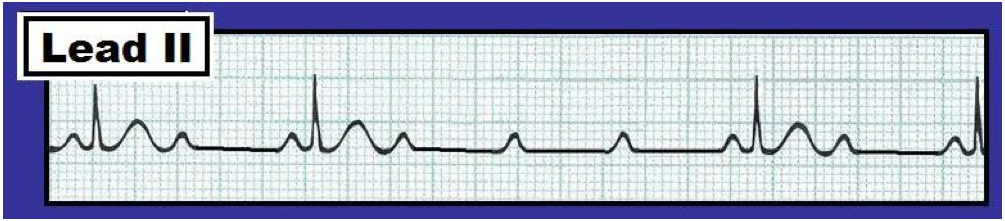
The lead II rhythm strip shown below in Figure 1 was obtained from an asymptomatic middle-aged adult. Is there complete (3rd degree) AV Block?  How would you proceed clinically?
Figure 1 - Lead II rhythm strip from an asymptomatic adult.
Is there 3rd degree AV block?
(Figure reproduced from ACLS-2013-ePub ).
- Note - Enlarge by clicking on Figures - Right-Click to open in a separate window.
INTERPRETATION:  The ventricular rhythm in the Figure is regular at a rate just over 50/minute (since the R-R interval is slightly less than 6 large boxes in duration).  The QRS complex is narrow, indicating a supraventricular etiology.  P waves are present – however, they are not consistently conducting.  Instead, the PR interval is changing. The PR interval preceding beats #3 and 4 (arrows) is clearly too short to conduct.
------------------------------------------------------
In ECG Blog #19 – we defined the 3 degrees of AV block as follows:
  • 1st degree AV block – in which all atrial impulses are conducted to the ventricles, albeit with delay (so that the PR interval exceeds 0.20 second).
  • 2nd degree AV block – in which some (but not all) atrial impulses are conducted to the ventricles.
  • 3rd degree (or “complete”) AV block – in which none of the atrial impulses are conducted to the ventricles, despite having adequate opportunity for conduction to occur.
The key to the diagnosis of complete AV block is in the last part of the definition: No atrial impulses are conducted to the ventricles “despite having adequate opportunity for conduction to occur”. Although beats #3 and 4 in Figure 1 (and possibly also beat #2) are not conducted to the ventricles – none of these beats has a “chance” to conduct, since the PR interval is simply too short. Thus, we have no idea if any degree of AV block is present – since we cannot tell from Figure 1 if P waves could conduct were they given the opportunity to do so. We therefore interpret this tracing as showing “AV dissociation” – since at least some P waves are unrelated to the QRS complexes that follow them. The term AV dissociation should never be used as a “diagnosis” per se.  Instead – it is the result of the underlying rhythm on the tracing.  In this case – the underlying rhythm is sinus bradycardia at a rate of 50/minute (the P-P interval is precisely 6 large boxes in duration for each of the P waves on this tracing). AV dissociation occurs by “default”.  That is – due to the relatively slow sinus rate, a nodal rhythm (at ~52/minute) takes over. This rhythm variant is not uncommonly seen in otherwise healthy young adult individuals. It may well be that there is no degree of AV block present, and that normal conduction will resume whenever the sinus node speeds up to a normal rate.
--------------------------------------------------
Distinction Beween AV Dissociation and Complete AV Block:  In contrast to Figure 1 – the lead II rhythm shown in Figure 2 illustrates complete (3rd degree) AV block.  
Figure 2 - Lead II rhythm strip showing 3rd degree AV block.
(Figure reproduced from ACLS-2013-ePub - pg 101)
- Note - Enlarge by clicking on Figures - Right-Click to open in a separate window.
The atrial and ventricular rates in Figure 2 are both regular with no relation between P waves and neighboring QRS complexes. The QRS complex is wide, indicating AV block at the ventricular level.  But the main distinction between Figures 1 and 2 is that despite more than adequate opportunity to conduct – none of the P waves in Figure 2 are conducted. We would not expect the P wave hidden in the T wave of beat #3 to conduct (because it occurs in the ST segment and is probably in the absolute refractory period). Similarly – the P wave preceding beat #1 in Figure 2 might not conduct (because this PR interval is not overly long). But virtually all other P waves on this tracing occur at a place when there should have been more than ample opportunity for conduction to occur. The diagnosis of complete AV block is therefore established.
--------------------------------------------

  - See also ECG Blog Reviews #19, 20 – and Section 20.0 of our ACLS-2013-ePub -
-------------------------------------------- 

Monday, May 9, 2011

ECG Interpretation Review #20 (High-Grade AV Block - Mobitz I vs Mobitz II?)

The Lead II rhythm strip shown below in Figure 1 was obtained was obtained from a patient during cardiopulmonary resuscitation. It is a follow-up tracing to our ECG Blog Review #19. The patient was hypotensive at the time Figure 1 was recorded. Does the rhythm represent 2nd degree AV Block, Mobitz Type II? How to proceed clinically?

Figure 1 - Lead II rhythm strip from a patient in cardiac arrest.
Is this 2nd degree AV block, Mobitz Type II?


INTERPRETATION:  The ventricular rhythm in Figure 1 is slow and irregular. Nevertheless – the QRS complex is narrow, and the atrial rate is regular at ~115/minute. The P waves immediately preceding each QRS complex manifest a fixed (and normal) PR interval. Thus, these P waves are conducting. This means that the rhythm is not complete AV block.  Since there are many non-conducted P waves on the tracing – the rhythm must represent some form of high-grade 2nd degree AV block.
-----------------------------------------------------

As discussed in ECG Blog Review #19 – Second degree AV blocks are generally classified into one of 3 types:
  • Mobitz I (AV Wenckebach) – in which the PR interval progressively lengthens until a beat is dropped.  This is by far the most common form of 2nd degree AV block. 
  • Mobitz II – in which there is a constant PR interval for consecutively conducted beats until one or more beats are dropped.
  • 2-to-1 AV Block – in which every-other-P-wave is conducted. Because one never sees two consecutively conducted beats – you can not tell if the PR interval is lengthening or not.  As a result – it is impossible to know for sure whether this form of 2nd degree AV block represents Mobitz I or Mobitz II.

-----------------------------------------------------
Returning to this Case: There are features of both Mobitz I (AV Wenckebach) and Mobitz II on the tracing shown in Figure 1. In favor of Mobitz II is the low conduction ratio and high grade of AV block. No less than 3 P waves in a row are non-conducted in the middle of the tracing.  However, the QRS complex is narrow – which is highly unusual for Mobitz II.
  • Clinically – the importance of distinguishing AV Wenckebach (Mobitz I) from Mobitz II relates to the much better prognosis of Mobitz I, a generally better response to treatment with Atropine, and a much lower likelihood of needing a pacemaker. That said – it is impossible to be certain which form of 2nd degree AV block is present from the tracing in Figure 1 alone, since one never sees two P waves in a row that conduct.  Thus, one can not tell if the PR interval is progressively increasing until the point of non-conduction.  Although unusual for Mobitz I – more than one P wave in a row may be blocked on occasion with this conduction disturbance.  From a practical treatment perspective, however – distinguishing between Mobitz I and Mobitz II appears to be less important since a pacemaker may be needed in either case unless the high-grade degree of AV block improves.

-----------------------------------------------------
FINAL (Advanced) PEARL: Even though far fewer P waves are conducted in Figure 1 from this Blog post (above) compared to the rhythm strip showing 2-to-1 AV conduction in Figure 1 from ECG Blog Review #19 (We reproduce that 2:1 AV block tracing below) – the “degree” of AV block per se has not necessarily “worsened”. Why not?  HINT: Has the atrial rate changed in the two tracings?

Figure 1 from ECG Blog Review #19 - Lead II rhythm strip showing 2:1 AV block.


ANSWER: The atrial rate in Figure 1 of this Blog post (at the top of this page) is now faster (~115/minute) than it was when there was 2:1 AV conduction (Note the P-P interval in Figure 1 from Blog Review #19 = 3 large boxes = an atrial rate of 100/minute). Rather than “worsening” of the degree of AV block per se – the lower conduction rate may simply be the result of the faster atrial rate.  Treatment of 2nd degree AV block with atropine may at times be a double-edged sword. It may improve the situation (by facilitating conduction through the AV node) – but it can at times worsen the situation by increasing the atrial rate by an amount that may be too fast to conduct. Bottom Line: We suspect that Figure 1 in this Blog post (at the top of this page) represents high-grade 2nd degree AV block, Mobitz Type I – because the QRS is narrow, and Mobitz I is by far the most common form of 2nd degree AV block. Even though treatment with atropine was appropriate (since this drug is often effective for Mobitz I) – atropine paradoxically worsened the situation in this case, because it further increased the atrial rate. Temporary pacing is indicated given bradycardia with hypotension.

------------------------------


  - See also ECG Blog #19 - and Section 20.0 in our ACLS-2013-ePub -
------------------------------

Sunday, May 8, 2011

ECG Interpretation Review #19 (2-to-1 AV Block - Mobitz I vs Mobitz II?)

The Lead II rhythm strip shown below was obtained from a patient during cardiopulmonary resuscitation. Systolic BP was 90 mmHg at the time this tracing was recorded.  Does this represent 2nd degree AV Block, Mobitz Type II?  How to proceed clinically?

Figure 1 - Lead II rhythm strip from a patient in cardiac arrest.
Is this 2nd degree AV block, Mobitz Type II?


INTERPRETATION:  The rhythm in the Figure 1 is slow but regular. The QRS complex is narrow, and a regular atrial rate is seen at 100/minute (arrows). Every other P wave conducts (as evidenced by the fact that a P wave does precede each QRS complex with a fixed PR interval! ).  Traditionally – the AV blocks are divided into 3 degrees based on severity of the conduction disturbance:
  • 1st degree AV block – in which all atrial impulses are conducted to the ventricles, albeit with delay (so that the PR interval exceeds 0.20 second).
  • 2nd degree AV block – in which some (but not all) atrial impulses are conducted to the ventricles.
  • 3rd degree (or “complete”) AV block – in which none of the atrial impulses are conducted to the ventricles, despite having more than adequate opportunity for conduction to occur.
====================================
Second degree AV blocks are further classified into 3 types (Figure 2):

Figure 2 - Types of 2nd Degree AV Block:
A) Mobitz I (AV Wenckebach); B) Mobitz II; and C) 2-to-1 AV Block.

  • Panel AMobitz I (AV Wenckebach) – in which the PR interval progressively lengthens until a beat is dropped.  This is by far the most common form of 2nd degree AV block. Mobitz I usually occurs at the level of the AV node.  As a result – the QRS complex is typically narrow. Mobitz I is generally associated with inferior infarction; it often spontaneously resolves, and typically responds to atropine (which works on the AV node).
  • Panel B = Mobitz II – in which there is a constant PR interval for consecutively conducted beats until one or more beats are dropped. Because Mobitz II typically occurs low down in the conduction system – the QRS complex is generally wide. This less common form of 2nd degree AV block is generally associated with anterior infarction; it usually does not respond to atropine – and is important to recognize because pacing will probably be needed.
  • Panel C = 2-to-1 AV Block – in which one never sees two consecutively conducted beats, so that you can not tell if the PR interval is lengthening or not.  As a result – it is impossible to know for sure whether this form of 2nd degree AV block represents Mobitz I or Mobitz II.

------------------------------------------------------
Returning to this Case: The rhythm in Figure 1 is the same as the one shown in Panel C of Figure 2. Because two conducted beats never occur in a row - we are unable to be certain of the type of 2nd degree AV block. That said  We strongly suspect this rhythm represents 2nd degree AV block, Mobitz Type I (AV Wenckebach) because: i) Mobitz I is so much more common than Mobitz II; and ii) the QRS complex is narrow, as it almost always is with Mobitz I.  Finding additional rhythm strips on this patient that clearly showed progressive lengthening of consecutively conducted QRS complexes before dropping a beat would strongly support our suspicion.  Clinically the distinction is important because no treatment (other than perhaps atropine) is likely to be needed for Mobitz I (especially given that the ventricular rate in Figure 1 is not overly slow at 50/minute). In contrast – pacing would probably be needed if the rhythm was Mobitz II.

-------------------------------------------------


   See also this pdf from Section 20.0 of our ACLS-2013-ePub 
-------------------------------------------------