
The simultaneously
recorded 2-lead rhythm strip shown below in Figure 1 was obtained from a young adult feeling “skips”.
- What is the rhythm?
- Is there intermittent AV block?
 |
| Figure 1: ECG from a young adult with “skips”. |
------------------------------------------
Interpretation of Figure 1:
The challenge in this tracing is to
find 2 normal beats in a row. The only
place where this seems to occur is at the very end of the rhythm strip for
beats #14,15. This tells us that the underlying rhythm is sinus tachycardia.
- Note how smooth the T wave is in both lead I and lead II for beats #14 and 15.
- Careful inspection of all T waves in both leads of this tracing reveals slight-but-real notching in almost all complexes. This is subtle. Be sure to click on the figure to enlarge the rhythm strip. Look first at lead I. Other than the T wave for beats # 8, 14 and 15 — there is a tiny but unmistakable notch in all other T waves.
- Each notch represents a PAC.
- Sinus P waves on this tracing are beats #1,3,4,5,7,9,10,11,12,14 and 15.
- The rhythm is atrial bigeminy — in that a PAC occurs in the T wave after each sinus beat (red arrows in Figure 2).
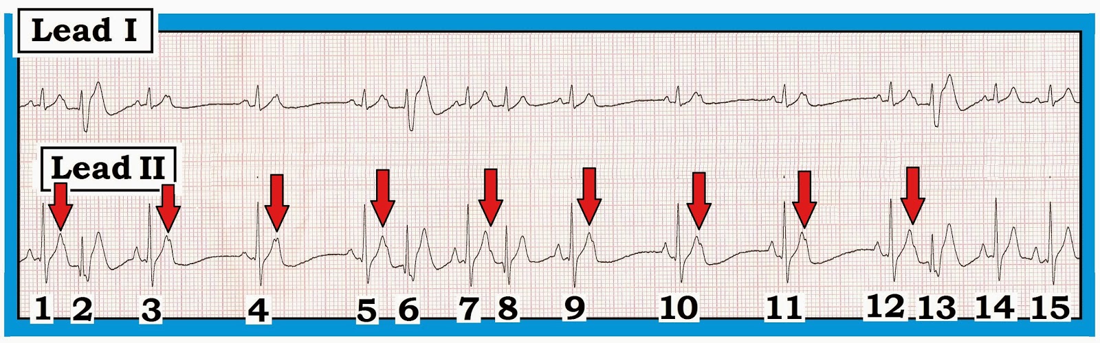 |
| Figure-2: Red arrows in the T wave of sinus beats indicates PACs. The rhythm is atrial bigeminy. Some PACs are conducted with aberration — others are blocked (See text). |
------------------------------------------
Looking
Closer: The KEY to interpreting this rhythm is to
appreciate the overall pattern. Once you identify 2 normal beats in a row (ie, beats #14,15) — you can determine what
the “normal T wave” should look like (= smooth
without any notching).
- Beat #1 is sinus.
- Beat #2 occurs early. The unmistakable notch in the T wave of beat #1 indicates that this is a PAC (Premature Atrial Contraction) and not a premature ventricular beat.
- The reason for the different and wider QRS morphology of beat #2 is that this PAC is conducted with aberration. Most aberrant beats conduct with a recognizable pattern of some form of bundle branch block or hemiblock, due to refractoriness of some portion of the conduction system. The deep and wide S wave of beat #2 in lead I with qR pattern in lead II suggests RBBB/LPHB aberration.
- Beat #3 is sinus.
- Note notching in the T wave of beat #3. This is the result of a blocked PAC (the PAC occurs so early in the refractory period that the entire conduction system is refractory, and the PAC is non conducted).
- Beat #4 is sinus. Another blocked PAC is hiding in (and notching) the T wave of beat #4.
- Beat #5 is sinus.
- Beat #6 is an aberrantly conducted PAC.
- Beat #7 is sinus.
- Beat #8 is an aberrantly conducted PAC.
- Beats #9,10,11 and 12 are sinus. Blocked PACs notch the T waves of beats #9,10,11.
- The PAC notching the T wave of beat #12 is conducted with aberrancy ( = beat #13).
- Beats #14 and 15 are the only two sinus beats seen to occur in a row on this tracing.
Bottom
Line: The rhythm
is sinus tachycardia with Atrial Bigeminy. PACs are either blocked or conducted
with aberration. There is no AV block. Given that the patient is a young adult
feeling “skips” — one should inquire about potential causative factors (ie, caffeine or other stimulants) that may
be causative.
-------------------------------------
- Acknowledgement: My appreciation to Simon Mortensen (of Odense, Denmark) for allowing me to use this case.
-------------------------------------
- For more
information — GO TO:
- See ECG Blog #14 — ECG Blog #15 — and — ECG Blog #33 for review of aberrant conduction.
- CLICK HERE — to download a pdf on Aberrant Conduction from Section 19.0 (in our ACS-2013-ePub).
No comments:
Post a Comment