NOTE: SEE BELOW for today’s ECG Media Pearl — which is an audio message relating to the phenomenon of deWinter-like T waves.
======================================
The patient whose ECG is shown in Figure-1 is a middle-aged man who presented to the ED with new-onset chest pain.
Which 2 of the following 5 choices are most correct?
- Extra Credit: WHY are the remaining 3 choices either less correct or wrong?
CHOICES to Consider:
- Choice A: The T waves in anterior leads may reflect a repolarization variant.
- Choice B: The anterior T waves and inferior ST-T waves suggest possible ischemia.
- Choice C: The cath lab should be immediately activated with an “OMI alert”.
- Choice D: The “classical” deWinter T wave pattern is present in the anterior leads.
- Choice E: Although a “classical” deWinter T wave pattern is not seen in ECG #1 — clinical implications are essential the same as if one was present.
 |
| Figure-1: ECG obtained from a middle-aged man with new chest pain (See text). |
MY THOUGHTS on ECG #1:
As always — it BEST to begin by systematic assessment of the ECG in question before moving on to clinical implications.
- NOTE: It could be easy to overlook that the rhythm in ECG #1 is not regular if one didn’t start by reviewing the long lead II rhythm strip at the bottom of the tracing. The mechanism of the rhythm is sinus — and all QRS complexes are preceded by similar-looking P waves with a constant PR interval. Variation in the R-R interval during the first half of the tracing is consistent with sinus arrhythmia — which then regularizes to normal sinus rhythm at ~80-85/minute toward the end of the rhythm strip.
- All intervals (PR, QRS, QTc) and the axis are normal. There is no chamber enlargement.
Regarding Q-R-S-T Changes:
- There are no Q waves.
- The R wave in anterior leads V1, V2, V3 is a bit slow to develop until transition occurs between V3-to-V4 (but there are no anterior Q waves or QS complexes).
- The most remarkable finding is the presence of hyperacute T waves that are seen in leads V2, V3 and V4. This is most marked in lead V3 (especially given relatively small S wave amplitude in this lead) — albeit there is no ST elevation in this lead. T waves in these 3 chest leads are clearly taller-than-they-should-be and overly “voluminous” (ie, fatter-at-their-peak and wider-at-their-base than would be expected given QRS amplitude in each respective lead).
- There is slight ST elevation in leads V1 and V2.
- Of note — there is subtle-but-definite J-point ST depression in leads V4, V5 and V6.
- There is some ST segment scooping, with slight depression in the inferior leads (including terminal T wave positivity after the ST-T depression).
Clinical IMPRESSION of ECG #1: This is not a repolarization variant (Choice A). Much more than “possible ischemia” (Choice B) — there are definite ECG abnormalities on this tracing in this patient with new-onsetchest pain. Given this history — ECG #1 should be interpreted as acute proximal LAD OMI (Occlusion-based MI) until proven otherwise — and the cath lab should be immediately activated (Choice C).
- PEARL #1: There are a number of findings in ECG #1 that suggest LAD occlusion is at a proximal location in this vessel. These include: i) ST elevation begins as early as in lead V1 — with hyperacute T waves also beginning early (in lead V2); and, ii) Reciprocal ST-T wave changes are seen in the inferior leads. Proximal LAD occlusion is usually also associated with at least slight ST elevation in lead aVL — but that is not present here.
- PEARL #2: Did YOU notice the lack of R wave progression until lead V4? The presence of a tiny-but-real initial r wave in leads V1 and V2 suggests that the septum remains intact — but lack of any increase in R wave amplitude until lead V4 may be indication of ongoing anterior infarction (ie, loss of anterior forces).
DeWinter T Waves:
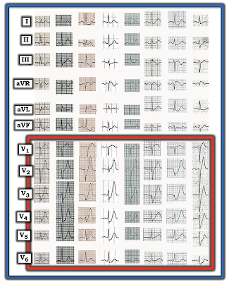
In 2008 — Robert J. deWinter and colleagues (Drs. Verouden, Wellens, and Wilde) submitted a Letter to the Editor to the New England Journal of Medicine (N Engl J Med 359:2071-2073, 2008) — in which they described a “new ECG pattern” without ST elevation that signifies acute occlusion of the proximal LAD (Left Anterior Descending) coronary artery.
- The authors recognized this pattern in 30 of 1532 (~2%) patients with acute anterior MI. Cardiac cath confirmed LAD occlusion in all cases — with ~50% of patients having a "wraparound" LAD. Left mainstem occlusion was not present.
- This was the authors’ original description of the new ECG pattern: “Instead of the signature ST-segment elevation — the ST segment showed 1-3 mm of upsloping ST depression at the J point in leads V1-to-V6 — that continued into tall, positive symmetrical T waves”.
- The QRS complex was usually not widened (or no more than minimally widened).
- Most patients also manifested 1-2 mm of ST elevation in lead aVR.
- NOTE: I’ve adapted Figure-2 from the original de Winter manuscript, published in this 2008 NEJM citation.
In their original 2008 manuscript — de Winter et al went on to describe the following additional features:
- “Although tall, symmetrical T waves have been recognized as a transient early feature that changes into overt ST elevation in the precordial leads — in this group of patients, this new pattern was static, persisting from time of the 1st ECG until the pre-cath ECG.”
- Hyperkalemia was not a contributing factor to this ECG pattern (ie, Serum K+ levels on admission were normal for these patients).
NOTE: Technically speaking — the de Winter T wave pattern as described in 2008 by de Winter et al differs from the finding of simple “hyperacute” anterior T waves — because ECG findings with a strict de Winter T wave pattern persist for an hour or more until the “culprit” LAD vessel has been reperfused.
- As I note above (and as illustrated in the example ECGs taken from the de Winter manuscript that are shown in Figure-2) — there should be involvement in all 6 chest leads with the strict de Winter pattern, with most leads showing several mm of upsloping J-point ST depression and giant T waves.
MY Observations regarding De Winter T Waves:
Over the past decade — I have observed literally hundreds of cases in numerous international ECG-internet Forums of deWinter-like T waves in patients with new cardiac symptoms.
- Many (most) of these cases do not fit strict definition of “de Winter T waves” — in that fewer than all 6 chest leads may be involved — J-point ST depression is often minimal (if present at all) in many of the chest leads — and, giant T waves are limited.
- ECG changes in many of these cases are not “static” until reperfusion, as was initially reported in 2008 by de Winter et al. Nevertheless, cath follow-up routinely confirms LAD occlusion.
- MY "Take": I believe there is a spectrum of ECG findings, that in the setting of new-onset cardiac symptoms is predictive of acute LAD occlusion as the cause. What will be seen on the ECG depends greatly on when during the process the ECG was obtained. While many of these patients do not manifest “true de Winter T waves” (because their ECG pattern does not remain static until reperfusion by coronary angioplasty) — for the practical purpose of promptly recognizing acute OMI — I don’t feel ( = my opinion) that it matters whether a “true” de Winter T wave pattern vs simple “hyperacute” T waves (that are deWinter-like) is present.
=====================================
NOTE: SEE BELOW for today’s ECG Media Pearl (#1) — which is an audio message relating to the phenomenon of deWinter-like T waves.
=====================================
Regarding Today’s Case: Comparing the chest lead ST-T wave appearance in ECG #1 with the 8 example tracings from the original de Winter et al manuscript shown in Figure-2 — ECG #1 is lacking in several features:
- Hyperacute T waves are not nearly as prominent in ECG #1 as in the 8 examples shown in Figure-2.
- The chest lead in ECG #1 that shows the most prominent T wave ( = lead V3) does not show any J-point ST depression.
- Only a single lead in ECG #1 shows both J-point ST depression and a hyperacute T wave ( = lead V4).
- BOTTOM LINE: ECG #1 does not satisfy criteria for a “STEMI” — because there is not enough ST elevation. It does not quite meet the original 2008 description for de Winter T waves. That said — it should still be obvious in this patient with new-onset chest pain that acute proximal LAD occlusion must be assumed until proven otherwise!
The 2nd ECG in Today’s Case:
A follow-up ECG was obtained about 3 hours after ECG #1. For purposes of comparison — I have put this ECG #2 under the initial tracing in Figure-3.
- How would YOU describe the ECG changes that have occurred since the initial tracing?
 |
| Figure-3: Comparison of the initial ECG in today’s case — with the follow-up ECG done about 3 hours later (See text). |
MY THOUGHTS on the Follow-Up Tracing:
ECG #2 shows that there has been extensive evolution during the 3 hours since the initial tracing was done:
- A significant Q wave is now seen in lead aVL of ECG #2, with suggestion of a tiny q wave in lead I. This is associated with loss of R wave amplitude (compared to ECG #1) in both of these high lateral leads.
- The poor R wave progression in leads V1, V2, and V3 persists — with loss of this initial r wave in lead V4 — leading to prominent Q waves in leads V4 and V5.
- There is now frank ST elevation in multiple leads, including leads I, aVL and V2-thru-V6.
- Inferior lead reciprocal changes of ST depression have deepened since ECG #1.
- Cardiac Cath was performed after ECG #2. It confirmed acute proximal LAD occlusion. Underlying multi-vessel disease (with ~50-60% stenoses) was present.
In CONCLUSION: Based on my experience over the past decade in commenting on hundreds of ECGs posted on various ECG internet forums — there are many “variants” of the original de Winter T wave pattern.
- Often, there won’t be J-point ST depression in anterior leads that also manifest overly large positive T waves — and, “deWinter-like” T wave patterns such as the one in today’s case may not necessarily persist until coronary reperfusion is accomplished.
- That said — despite not quite satisfying strict criteria for the de Winter T wave pattern described in the original 2008 NEJM manuscript — clinical implications of the findings in ECG #1 are essentially the same as if those strict criteria were present = acute proximal LAD occlusion until proven otherwise (Choice E).
===================================
- Acknowledgment: My appreciation to 유영준 (from Seoul, Korea) for the case and this tracing.
===================================
| TODAY’s ECG Media PEARL #1 (3:00 minutes Audio): — relates to the phenomenon of deWinter-like T waves. |


Nice description and beautifully explained
ReplyDeleteTHANK YOU! — :)
DeleteExcellent teaching ECG like always.
ReplyDeleteMany thanks for posting so often in 2021 !
Gilles Mugnier
Merci bien Gilles! — :)
DeleteLove the new audio pearl !
ReplyDeleteThank you! — :)
Delete