
- YOUR thoughts on this case?
- HINT: The optimal answer will address both the rhythm diagnosis and clinical management.
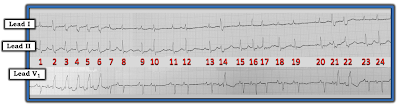 |
| Figure-1: ECG obtained from an elderly woman awaiting surgery. |
=======================================
NOTE #1: Some readers may prefer at this point to listen to the 8:00 minute ECG Audio PEARL before reading My Thoughts regarding the ECG in Figure-1. Feel free at any time to review to My Thoughts on this tracing (that appear below ECG MP-29).
=======================================

Today’s ECG Media PEARL #29 (8:00 minutes Audio) — Reviews WHAT the Ashman Phenomenon is — HOW to use it clinically? — and — whether the Ashman phenomenon is accurate when the underlying rhythm is AFib?
- NOTE #2: For detailed review of the Ashman Phenomenon — with illustration of its clinical application — Please See ECG Blog #70. Use of the Ashman phenomenon with AFib is reviewed in ECG Blog #71.
- NOTE #3: Additional material relevant to today's case can be found below in the Addendum.
- The first priority in assessing any acute arrhythmia — is to ensure that the patient is hemodynamically stable. Once you have done so — then by definition, you have at least a moment of time to interpret the rhythm.
- In my experience — the most accurate and time-efficient way to interpret any cardiac arrhythmia — is to use a Systematic Approach. As emphasized often on this ECG Blog — I favor the Ps, Qs, 3R Approach (See ECG Blog #185).
- PEARL #1: In addition to using a systematic approach and using calipers — I find it extremely helpful to look for an underlying rhythm. Simply stated — when there is more than one active feature in the rhythm — LOOK FIRST for the EASIER elements to interpret. Save the more difficult elements for later. For example, in Figure-1 — I would save the run of wider, faster beats (ie, beats #2-thru-6) until after I’ve assessed other parts of this tracing that look to be easier to interpret.
- Ignoring the wider beats for the time being (as per Pearl #1) — the QRS in all 3 leads is narrow for the remaining beats ( = beats #1; 7-13; 15-20; and 23, 24). Therefore — the underlying rhythm is supraventricular.
- This underlying rhythm for these narrow beats is not Regular. As a result, the Rate varies — but a tachycardia is present, as the R-R interval is virtually always less than 3 large boxes in duration (ie, the average rate is clearly over 100/minute).
- P waves are present — and they do appear to be Related to neighboring QRS complexes, in that a P wave precedes virtually all QRS complexes.
- PEARL #2: P wave morphology appears to be changing, almost from beat-to-beat thoughout the entire rhythm strip in Figure-1. This constantly changing P wave morphology is subtle-but-real, although not easy to appreciate from lead V1 — because most P waves in this lead are pointed. The KEYS to appreciating the variation in P wave morphology are to: i) Note that the PR interval does not remain the same; and, ii) Look at P waves in each of the simultaneously-recorded leads. For example, although the P wave preceding beats #11, 12 and 13 in lead V1 is pointed at its peak — the change in morphology from one P wave to the next preceding these same beats #11, 12 and 13 is much more obvious in simultaneously-recorded leads I and II.
- Looking closely — the P waves preceding beats #14, 21 and 24 in lead V1 appear to be biphasic. While true that it sometimes is difficult to distinguish between variations in P wave morphology that result from baseline artifact or the "normal" variation in shape than can often be seen between one sinus P wave and the next — I interpreted the above described variation in P wave morphology as real.
- MAT is not a common diagnosis. As a result, in order to differentiate MAT from the much more commonly encountered irregularly irregular rhythm (which is AFib) — we need to be certain we are seeing multiple different P wave morphologies that are constantly changing. Pearl #2 above describes how we are!
- PEARL #3: The diagnosis of MAT almost always occurs in one of 2 common predisposing settings. These 2 settings are: i) In patients with severe, often longstanding pulmonary disease; and/or, ii) In acutely ill patients with multi-system disease (ie, sepsis, shock, electrolyte and/or acid-base disorders). Awareness of this association is clinically important — because success in treating MAT depends on identifying and correcting this underlying cause!
- PEARL #4: There is a phenomenon I've described as "Birds of a Feather". By this I mean that if there are multiple instances of an event in an arrhythmia (such as multiple early beats) — and we know that the overwhelming majority of these early beats are PACs, but we are uncertain about the etiology of a few widened early beats — "Think birds of a feather flock together" — such that rather than postulating an additional etiology for these few widened beats (such as PVCs) — it becomes likely that these unknown early beats are also PACs that are widened because of aberrant conduction (See Audio Pearl MP-67 below in the Addendum for more on the "Birds of a Feather" phenomenon).
- The first wide beat in each of the 3 places where wide beats occur on the long lead rhythm strip is clearly preceded by a premature P wave (RED arrows before beats #2, 14 and 21 in Figure-2).
- P waves also precede the other wide beats on this rhythm strip (PINK arrows before beats #3, 4, 5, 6 and 22).
- Each of the widened beats in Figure-2 manifest a RBBB (Right Bundle Branch Block) morphology, with upright widened QRS in lead V1 and wide, terminal S wave in lateral lead I. The first wide beat in each grouping (ie, beats #2, 14 and 21) manifests the highly specific triphasic rsR' pattern that is so characteristic of aberrant conduction (See Video Pearl MP-28 below in the Addendum for full explanation why RBBB morphology is the most common pattern with aberrant conduction).
- PEARL #5: The Ashman Phenomenon is present, and explains why beats #14, 21 and 22 conduct with RBBB aberration. As explained in detail in Audio Pearl MP-29 near the top of this page — the preceding R-R interval is proportional to the length of the subsequent relative refractory period. As a result — aberrantly-conducted beats often follow the longest pauses in a rhythm strip (ie, The longest pauses in Figure-2 = the R-R intervals between beats #12-13, and between beats #19-20).
- Beyond-the-Core: Did YOU notice that in addition to the typical RBBB morphology for the widened beats — that a typical hemiblock morphology is also present for most of the widened beats? Specifically — the deep S wave descent in lead I for beats #3, 4, 5 and 6 is characteristic of LPHB (Left Posterior HemiBlock) conduction — and predominant negativity of the QRS in lead II for beats #14 and 21 is characteristic of LAHB (Left Anterior HemiBlock) conduction. The presence of this alternating typical pattern of bifascicular block conduction (RBBB/LPHB and RBBB/LAHB) provides yet one more finding in strong support of aberrant conduction.
 |
| Figure-2: I have labeled P waves before each of the widened beats in Figure-1 (See text). |
- As emphasized above in Pearl #3 — the success in treating MAT depends on identifying and correcting the underlying cause of this rhythm. Awareness that the elderly patient in today's case had both hypokalemia and hypoxemia explains why treatment with several antiarrhythmic agents failed to control her arrhythmia.
- In addition to the underlying rhythm of MAT — the rapid succession of PACs at the beginning of this rhythm strip (ie, beats #1-thru-6) constitues a run of Atrial Tachycardia, in which beats #2-thru-6 are wide because of aberrant conduction. There is no evidence of VT on this tracing. Hopefully, correction of the electrolyte disturbance and the patient's hypoxemia will help to resolve both the MAT and the runs of Atrial Tachycardia.
- See ECG Blog #185 — for review of the Systematic Ps, Qs, 3R Approach to rhythm interpretation.
- See ECG Blog #70 — for Review of the Ashman Phenomenon.
- See ECG Blog #71 — for Review of why the Ashman Phenomenon may be less reliable with AFib (also reviews typical RBBB aberrancy morphology).
- See ECG Blog #212 — for Step-by-Step discussion of another case with the Ashman Phenomenon.
- See ECG Blog #211 — Reviews WHY some early beats and some SVT rhythms are conducted with Aberration (and why the most common form of aberrant conduction manifests RBBB morphology).
- See ECG Blog #65 — for Review of MAT (and how this rhythm has similar clinical implications as for sinus rhythm with multiple PACs).
- See ECG Blog #199 — for a thorough Review of MAT (with brief Video Pearl to facilitate recognition of this entity).
- See ECG Blog #253 — Reviews a case with multiple aberrantly-conducted beats.
Relevant ECG Blog Posts to Today’s Case:
- ECG Blog #140 — Example of alternating Bifascicular Block Aberration.
- ECG Blog #14 — Example of Blocked PACs.
- ECG Blog #15 — Example of a WCT due to Aberrant Conduction.
- ECG Blog #33 — Example of PACs with varying degrees of Aberrant Conduction.
- mm
- ECG Blog #155 — What is a Wandering Pacemaker?
- ECG Blog #200 — More on a Wandering Pacemaker (including brief Audio Pearl on this topic).
Today’s ECG Media PEARL #28 (4:45 minutes Video) — Reviews WHY some early beats and some SVT rhythms are conducted with Aberration (and why the most common form of aberrant conduction manifests RBBB morphology).
- NOTE #3: I have excerpted a 6-page written summary regarding Aberrant Conduction from my ACLS-2013-ePub. This appears below in the Addendum (in Figures-4, -5, and -6).
- CLICK HERE — to download a PDF of this 6-page file on Aberrant Conduction.
=======================================

- See ECG Blog #253 — for review of a case illustrating application of the "Birds of a Feather" principle.
=================================
For a Summary of KEY points related to MAT:
 |
| Figure-3: Summary of KEY points related to MAT. |
=================================
In the following 3 Figures — I post written summary from my ACLS-2013-ePub regarding the basics of Aberrant Conduction.
- CLICK HERE — for a PDF of this 6-page file on the basics of Aberrant Conduction that appears in Figures-4, -5, -6.
 |
| Figure-4: Aberrant Conduction — Refractory periods/Coupling intervals (from my ACLS-2013-ePub). |
 |
| Figure-5: Aberrant Conduction (Continued) — QRS morphology/Rabbit Ears. |
 |
| Figure-6: Aberrant Conduction (Continued) — Example/Summary. |
No comments:
Post a Comment