Saturday, November 25, 2023

ECG Video Blog #405 — Is AV Block Complete (vs AV Dissociation)


======================================
 CLICK HERE — for a Video presentation of this case! (19:40 min.)

  • Below are slides used in my video presentation.
  • For full discussion of this case — See ECG Blog #191 

======================================


The 2-lead rhythm strip shown in Figure-1 was obtained from an elderly woman who presented to the ED following a syncopal episode. On the basis of this rhythm strip — she was diagnosed as being in complete AV Block.

  • Question #1Is there AV Dissociation in Figure-1?
  • Question #2: Do YOU agree that the rhythm shown in this figure represents complete ( = 3rd-degree) AV Block?
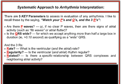
Figure-1: Is this complete AV Block


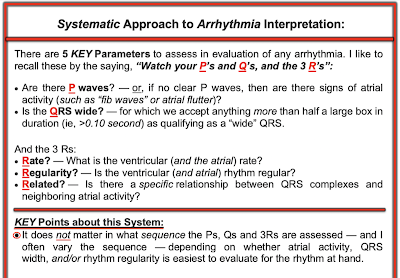
Figure: The Ps, Qs, 3R Approach to Rhythm Interpretation.


Figure: It does not matter in what sequence you assess the 5 Parameters (and I often change the sequence, depending on the rhythm ... ).

=======================================================


Figure: If the AV block is neither 1st-degree nor 3rd-degree — then it will be some type of 2nd-degree AV Block!


Figure: 1st-degree AV Block — is simply a sinus rhythm with a long PR interval (ie, more than 1 large box in duration = >0.20 second). It is EASY to diagnose.


Figure: 3rd-degree AV block with ventricular escape. 3rd-degree AV Block is usually also EASY to diagnose — because most of the time the ventricular "escape" rhythm will be regular (or almost regular) — in contrast to conducted beats that will often occur earlier-than-expected ...


Figure: The 3 types of 2nd-degree AV blockPanel A: Mobitz I 2nd degree AV block with gradual prolongation of the PR interval until a P wave is droppedPanel B: Mobitz II with QRS widening and a fixed PR interval until sudden loss of conduction with successive nonconducted P wavesPanel C: 2nd-degree AV block with 2:1 AV conduction. It is impossible to be certain if 2:1 AV block represents Mobitz I or Mobitz II — because we never see 2 conducted P waves in a row — and, therefore can not tell if the PR interval would progressively lengthen prior to nonconduction IF given a chance to do so ... (That said — 90-95% of 2nd-degree AV Blocks are Mobitz I — and Mobitz I especially likely if the QRS is wide, with 1st-degree, in a patient with acute inferior MI).


Figure: 2nd-degree AV block with 2:1 AV conduction.


Figure: This is high-grade 2nd-degree AV Block — as identified by the finding of at least 2 consecutive on-time P waves that fail to conduct despite adequate opportunity to do so (occurring here between beats #2-3).


=======================================================


Figure: AV dissociation by "default" — because the SA node slows, with result takeover by the AV node. There is not necessarily any degree of AV block with this! 


Figure: AV dissociation by "usurpation" — because either the AV node or the ventricles speed up, and take over the rhythm from the SA node. There is not necessarily any degree of AV block with this! (and the KEY is to figure out WHY the AV node or ventricles have "taken over" the rhythm). 


Figure: Anatomic levels of 3rd-degree AV Block. Panel A: Complete AV Block at the ventricular level. There is a regular atrial rhythm — and the QRS is wide with an idioventricular escape rhythm at a rate between 20-40/minute. Panel B: Complete AV Block at a higher level (probably in the AV node) — as suggested by the presence of a narrow QRS escape rhythm at a rate between 40-60/minute.

NOTE: The KEY criterion for diagnosing complete AV block — is that none of the on-time sinus P waves are being conducted to the ventricles despite having more than adequate opportunity to do so! (which usually requires a long enough rhythm strip at a slow enough rate = <50-55/minute — in order to guarantee that on-time P waves are occurring at all points in the cycle — but still fail to conduct).


=======================================================


=======================================================


Figure: Laddergram of today's rhythm through the Atrial Tier.


Figure: Completed laddergram of today's rhythm. There is AV dissociation for the first 5 beats with ventricular escape (AV dissociation as a result of AV block). Because of underlying sinus arrhythmia — the P wave in front of beat #6 occurs at an opportune time, and is able to conduct to the ventricles. The remaining beats ( = beats #6-thru-9) conduct with 2:1 AV block. Because Mobitz I is some much more common than Mobitz II — and especially because the QRS complex for these conducted beats is narrow — there is almost certainly 2nd-degree AV Block of the Mobitz I Type ( = AV Wenckebach) — here with AV dissociation initially, and 2:1 AV block for the last few beats in the tracing.


==================================


Additional Material on Today's CASE:


ECG Media PEARL #4 (4:30 minutes Audio): — takes a brief look at the AV Blocks — and focuses on WHEN to suspect Mobitz I.


ECG Media Pearl #8 (8:20 minutes Video) — ECG Blog #191 — Distinguishing between ADissociation vs Complete AV Block (2/6/2021).


ECG Media Pearl #9 (5:40 minutes Video) — ECG Blog #192 — Reviews the 3 Causes of AV Dissociation (2/9/2021).


  • Section 2F (6 pages = the "short" Answer) from my ECG-2014 Pocket Brain book provides quick written review of the AV Blocks (This is a free download).
  • Section 20 (54 pages = the "long" Answer) from my ACLS-2013-Arrhythmias Expanded Version provides detailed discussion of WHAT the AV Blocks are — and what they are not(This is a free download). 


=======================

Related ECG Blog Posts to Today’s Case: 

  • ECG Blog #185 — Review of the Ps, Qs, 3R Approach for systematic rhythm interpretation.
  • ECG Blog #188 — Reviews how to read and draw Laddergrams (with LINKS to more than 50 laddergram cases — many with step-by-step sequential illustration).
  •  
  • ECG Blog #164 — Which reviews step-by-step the diagnosis of a Mobitz I 2nd-degree AV block (with sequential laddergram illustration).

  • ECG Blog #168 — A complex dual-level AV Wenckebach (Laddergram).

  • ECG Blog #154 and ECG Blog #55 and ECG Blog #224 and ECG Blog #232 — Acute MI with AV Wenckebach.

  • ECG Blog #63 — Mobitz I with Junctional Escape Beats.


No comments:

Post a Comment