
Today's CASE:
I was asked to interpret the ECG in Figure-1 — obtained from a 60-year old man with lung cancer. The patient had developed myocarditis as a complication of drug treatment for his disease. This was further complicated by recurrent VT.
- Some time after the VT resolved — the ECG in Figure-1 was recorded.
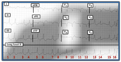 |
| Figure-1: ECG from a 60-year old man with lung cancer, myocarditis — and a history of prior recurrent VT. |
QUESTION:
- How would YOU interpret this ECG?
ANSWER:
I thought it might be most helpful to address today's case by means of an ECG Video, which I present below:
- NOTE: This is a complex tracing — and — I fully acknowledge that I am not certain about all aspects of the ECG in Figure-1. That said — I thought this case makes for excellent illustration of the thought process for working through a complex arrhythmia. It also serves to illustrate how sometimes the use of a Laddergram can suggest the etiology for an arrhythmia that otherwise might have been insolvable if a ladergram had not been used.
- To EMPHASIZE: I believe there is value in following this case regardless of how experienced you may or may not be with ECG interpretation. You do not have to know how to draw Laddergrams in order to be able to read them! This ECG Video will cover some important material for any level of interpreter.
ECG Media Pearl #82 (8:40 minutes Video) — Detailed analysis of this challenging arrhythmia (including ECG Video wth step-by-step analysis of my proposed Laddergram).
- CLICK HERE to download a PDF of all tracings shown in today's ECG Video (including step-by-step construction of my proposed Laddergram).
NOTE: Below in Figure-2 is my proposed Laddergram solution for today's case. I welcome input from readers — including additional proposed solutions for this case. To EMPHASIZE: This is not a simple case — and since I'm not certain of the full mechanism, my purpose in presenting this rhythm was not solely to find "the Answer". Instead — I'd be extremely happy if you:
- Recognized the overall long-short "pattern" to the rhythm (ie, "group" beating).
- Appreciate the use of the Ps, Qs, 3R Approach (Figure-3).
- See the importance of using all 12 leads of an ECG to help in determining the rhythm.
- Appreciate that P waves may be "related" to neighboring QRS complexes (and therefore conducting) — even when the relationship is not overly evident.
- Appreciate how a laddergram may suggest an etiology for a complex rhythm that otherwise might not be conceivable.
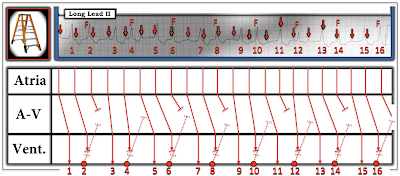 |
| Figure-2: My proposed Laddergram for today's case. Full discussion of my rationale for derivation of this Laddergram is covered in the above ECG Video (ECG-MP-82). |
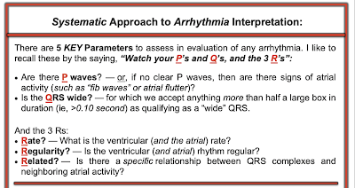 |
| Figure-3: The systematic Ps, Qs, 3R Approach for Rhythm Interpretation. |
=======================
Acknowledgment: My appreciation to Antranik Ohanian (from Beirut, Lebanon) for the case and this tracing.
=======================
=============================
Relevant LINKS to Today's Case:
=============================
How to Draw a Laddergram (Step-by-Step Demonstration)
- See ECG Blog #69 — for a verbal Step-by-Step on drawing a Laddergram.
- See ECG Blog #188 — for a brief ECG Video review on the basics of what a Laddergram is — with LINKS at the bottom of the page to more than 50 ECG blog posts in which I review illustrative laddergrams.
- See ECG Blog #164 — for a user-friendly rhythm-solving approach to AV Wenckebach, followed by Step-by-Step construction of the Laddergram.
- CLICK HERE — to DOWNLOAD my Free PowerPoint Laddergram STENCIL for your use as desired.
Additional Relevant Material to Today's Case:
- See ECG Blog #185 — for review of the Systematic Ps, Qs, 3R Approach to rhythm interpretation.
- See ECG Blog #128 — for review of the concept of Fusion Beats.
- See ECG Blog #129 — for more on Fusion Beats (with laddergram illustration).
===================================
No comments:
Post a Comment