
======================================
CLICK HERE — for a Video presentation of this case! (22:30 min.)
- Below are slides used in my video presentation.
- For full discussion of this case — See ECG Blog #292 —
======================================
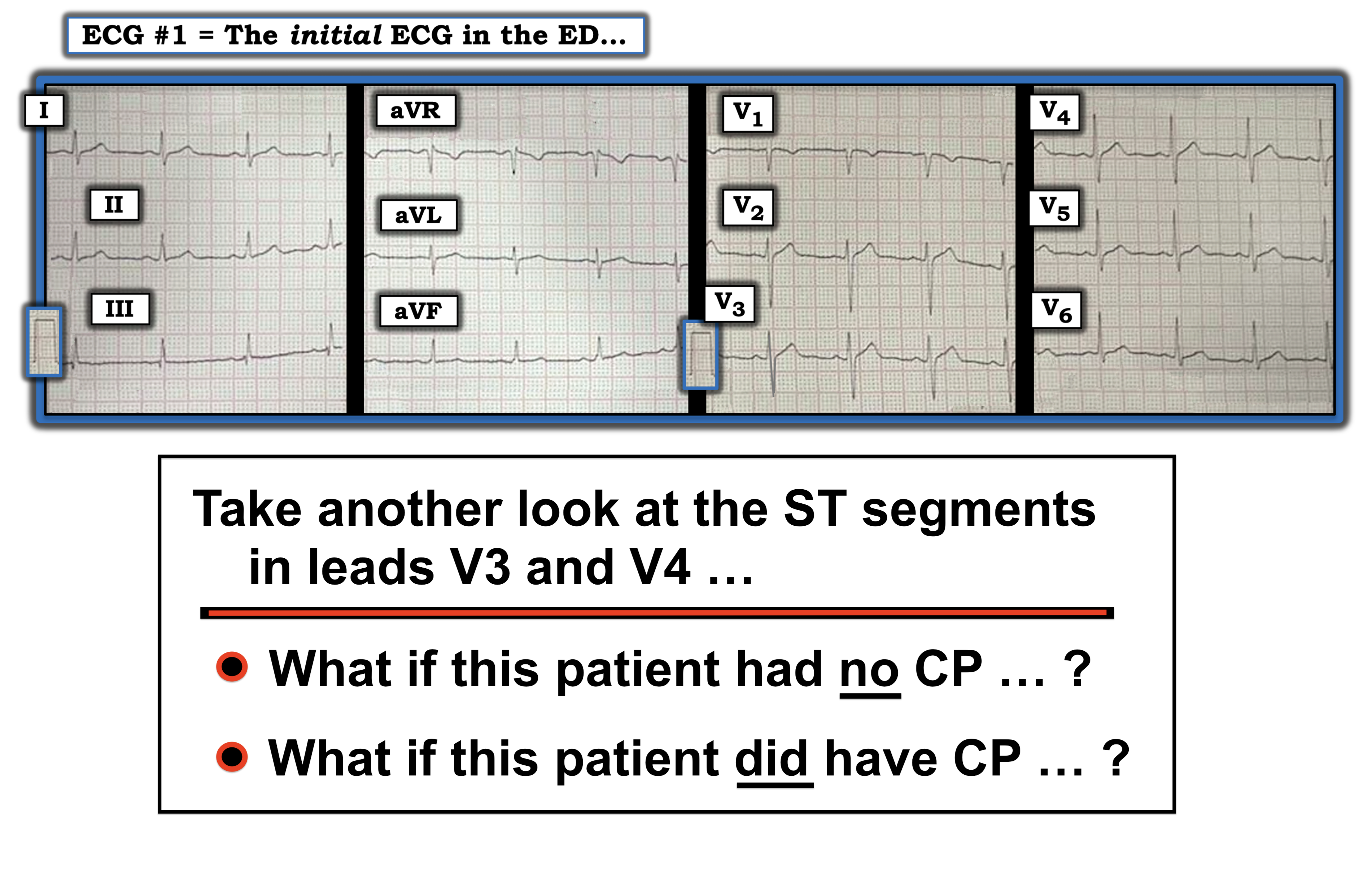
The 2 ECGs shown in Figure-1 were obtained from a man in his 30s — who presented to the ED (Emergency Department) with chest pain that began several hours earlier. ECG #2 was recorded 1 hour after ECG #1. Initial troponin was negative.
Cardiac cath was advised — but the patient refused. Instead, he left the hospital — only to be found dead at home 36 hours later.
- How would YOU interpret the serial ECGs shown in Figure-1?
- WHY did the patient die?
 |
| Figure-1: The 2 ECGs in today's case (See text). |
-USE.png)
-Why_Wide-USE%20copy%202.png)

-USE.png)


-USE.png)

-USE.png)
-USE.png)


=======================
ADDENDUM: Some additional material on ECG diagnosis of OMI.
=======================

ECG Media PEARL #39a (4:50 minutes Audio) — Reviews the concept of Dynamic ST-T Wave Changes (and how this ECG finding can assist in determining if acute cardiac cath is indicated).

ECG Media PEARL #46a (6:35 minutes Audio) — Reviews HOW to compare Serial ECGs (ie, Are you comparing "Apples with Apples" — or — with Oranges?).
Related ECG Blog Posts to Today’s Case:
- ECG Blog #205 — Reviews my Systematic Approach to 12-lead ECG Interpretation.
- ECG Blog #183 — Reviews the concept of deWinter T-Waves (with reproduction of the illustrative Figure from the original deWinter NEJM manuscript).
- ECG Blog #222 — Reviews the concept of Dynamic ST-T wave changes, in the context of a detailed clinical case.
- ECG Blog #260 — Reviews another case that illustrates the concept of "dynamic" ST-T wave changes.
- ECG Blog #218 — Reviews HOW to define a T wave as being Hyperacute?
- ECG Blog #230 — Reviews HOW to compare Serial ECGs (ie, "Are you comparing Apples with Apples or Oranges?").
- ECG Blog #193 — Reviews the concept of why the term “OMI” ( = Occlusion-based MI) should replace the more familiar term STEMI — and — reviews the basics on how to predict the "culprit" artery.
- ECG Blog #194 — Reviews how to tell IF the “culprit” (ie, acutely occluded) artery has reperfused using clinical and ECG data.
- ECG Blog #115 — Shows an example of how drastically the ECG may change in as little as 8 minutes.
- The January 9, 2019 post in Dr. Smith's ECG Blog (Please scroll down to the bottom of the page to see My Comment). This case is remarkable for the dynamic ST-T wave changes that are seen. It's helpful to appreciate: i) That acute ischemia/infarction is not the only potential cause of such changes (cardiac cath was normal); ii) That changes in heart rate, frontal plane axis and/or patient positioning can not always explain such changes; and, iii) That entities such as repolariztion variants, LVH and/or acute myopericarditis may all contribute on occasion to produce an evolution of challenging dynamic ST-T wave changes on serial ECGs.
- The August 22, 2020 post in Dr. Smith's ECG Blog — which illustrates another case of dynamic ST-T wave changes that resulted from a repolarization variant.
- The July 31, 2018 post in Dr. Smith's ECG Blog (Please scroll down to the bottom of the page to see My Comment). This case provides an excellent example of dynamic ST-T wave changes on serial tracings (that I illustrate in My Comment) in a patient with an ongoing acutely evolving infarction.
what about T wave changes in III and aVF?
ReplyDelete